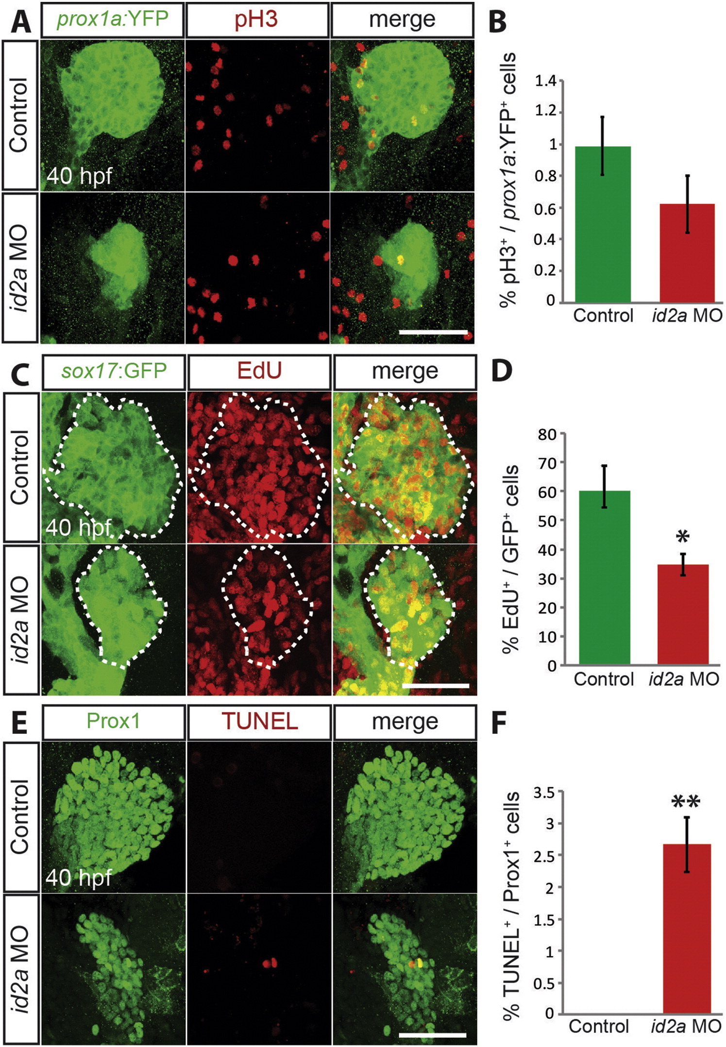
Fig. 4
id2a knockdown decreases hepatoblast proliferation and increases cell death in the developing liver. (A) Whole-mount immunostaining with anti-pH3 (red) anti-GFP(green) antibodies in Tg(prox1a:YFP) embryos. The total number of prox1a:YFP+ hepatic cells per liver is 316 ± 8.5 in controls and 156 ± 5.6 in id2a MO-injected embryos. (B) A graph showing the percentage of pH3+ cells among prox1a:YFP+ hepatic cells (n = 10). (C) EdU labeling (red), combined with anti-GFP immunostaining (green), in Tg(sox17:GFP) embryos reveals a significant reduction of proliferation in the liver of id2a MO-injected embryos at 40 hpf compared with controls. Dotted lines outline the liver. The total number of sox17:GFP+ cells per liver is 220 ± 16.6 in controls and 127 ± 12.7 in id2a MO-injected embryos. (D) A graph showing the percentage of EdU+ cells among GFP+ hepatoblasts (n = 10). (E) TUNEL labeling (red) combined with anti-Prox1 immunostaining (green) reveals apoptosis in the liver of id2a MO-injected embryos at 40 hpf, but not in controls. The total number of Prox1+ cells per liver is 276 ± 20.5 in controls and 140 ± 8.6 in id2a MO-injected embryos. (F) A graph showing the percentage of TUNEL+ cells among Prox1+ hepatoblasts (n = 10). *p < 0.05, **p < 0.005; error bars, ± s.e.m. Scale bars: 50 µm.
Reprinted from Mechanisms of Development, 138 Pt 3, Khaliq, M., Choi, T.Y., So, J., Shin, D., Id2a is required for hepatic outgrowth during liver development in zebrafish, 399-414, Copyright (2015) with permission from Elsevier. Full text @ Mech. Dev.

