
|
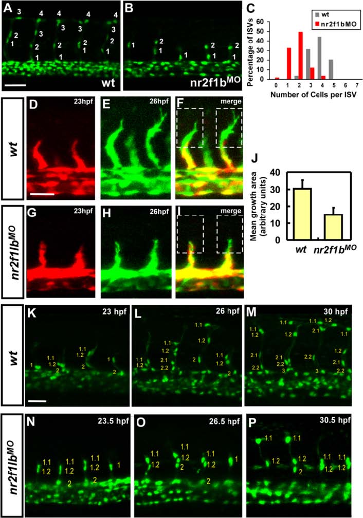
Fig. 5
Nr2f1b is required for the growth of intersegmental vessels. The number of cells forming each ISV were counted in wild type control Tg(fli1a:negfp) y7 (a) and nr2f1b morphant embryos (b) at 30 hpf. c Proportional distribution of ISVs containing 1-7 cells in both conditions. d-j Time-lapse imaging of wild type Tg(kdrl:eGFP) la116 (d-f) and nr2f1b morphant (g-i) embryos to examine the extension of ISV tip cell filopodia. Confocal images at 23 hpf (red; d, g) and 26hpf (green; e, h) were merged (f, i). The extension of tip cell was quantitated by pixel intensity and shows reduced extension of tip cell in nr2f1bMO (j). k-p Time-lapse imaging of wild type Tg(fli1a:nEGFP) y7 (k-m) and nr2f1b morphant (n-p). Scale bars represent 50 µm.