
|
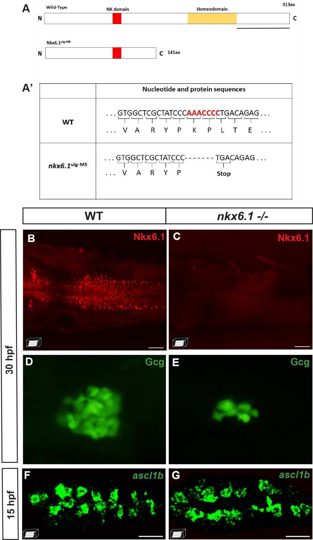
Fig. S4 ascl1b expression is not repressed by Nkx6.1. A Schematic representation of wild-type (WT) and mutant Nkx6.1 ulg-M5 proteins with the yellow box representing the homeodomain and the red box representing the NK domain. The coding region of the mutant Nkx6.1 ulg-M5 protein contains a 7-bp deletion after the aa 141 codon, leading to the apparition of a STOP codon just after the deletion. The black line represents the region of the protein recognized by the Nkx6.1 antibody. A′ Table showing part of the nucleotide and protein sequence of the nkx6.1 gene and of the mutated form in the Nkx6.1 ulg-M5 mutant. B, C Nkx6.1 immunodetection showing the loss of the full-length Nkx6.1 protein in the nkx6.1 -/- mutant. D, E Glucagon immunodetection showing a drastic reduction of the number of glucagon-expressing cells in the nkx6.1-/- mutant compared to the WT embryo at 30 hpf. This phenotype is the same as the one of embryos injected with the MO1 translation-blocking morpholino, targeting the translation start site of nkx6.1 mRNA [18], shown to prevent nkx6.1 expression efficiently in the neural tube [72]. This strongly suggests that the mutant Nkx6.1 ulg-M5 is a null mutant. F, G Confocal projection images of fluorescent WISH showing equivalent number of ascl1b+ cells in WT and nkx6.1-/- mutants at 15 hpf. All views are ventral with the anterior part to the left. Scale bars = 40 µM.