
Fig. 5
- ID
- ZDB-IMAGE-151030-16
- Antibodies
- Publication
- Miesfeld et al., 2015 - Yap and Taz regulate retinal pigment epithelial cell fate
- All Figures
- Figures for Miesfeld et al., 2015
|
Fig. 5
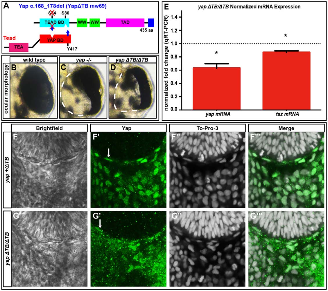
Tead-binding-deficient yapΔTB/ΔTB mutants lack RPE but maintain yap mRNA and Yap protein levels. (A) The Tead-binding-deficient yapΔTB/ΔTB zebrafish mutant. (B-D) 48hpf whole eyes showing that yap-/- and yapΔTB/ΔTB mutants lose a subset of RPE cells. Dashed lines indicate the border of the eye. (E) qRT-PCR analysis of whole embryos at 32hpf revealing a decrease in yap (1.6-fold, *P=0.0052) and taz (1.15-fold, *P=0.0038) mRNA in yapΔTB/ΔTB mutants. Dotted line indicates the normalized expression levels of yap and taz in wild-type embryos. An unpaired t-test was performed and statistical significance was determined using the Holm-Sidak method. Error bars indicate s.e.m. (F-G′′′) Yap protein expression in yap+/ΔTB (F-F′′′) and yapΔTB/ΔTB (G-G′′′) at 28hpf. Yap protein is present in flattened RPE nuclei (arrows) and periocular cells in yap+/ΔTB and yapΔTB/ΔTB embryos.