
Fig. 7
- ID
- ZDB-IMAGE-151022-7
- Genes
- Antibodies
- Publication
- Mateus et al., 2015 - Control of tissue growth by Yap relies on cell density and F-actin in zebrafish fin regeneration
- All Figures
- Figures for Mateus et al., 2015
|
Fig. 7
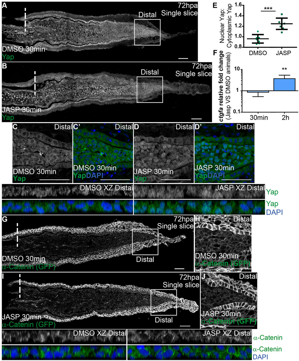
F-actin controls Yap intracellular dynamics. (A-D′) Representative immunofluorescence with anti-Yap antibody in 72hpa longitudinal sections of α-Catenin transgenics injected with jasplakinolide (JASP; B) and DMSO controls (A). (C-D′) High magnification images of the boxed areas in A,B showing Yap and DAPI in DMSO (C,C′) and JASP (D,D′) conditions. Corresponding xz projections of the distal blastemas shown in C,D highlight intracellular localization (DMSO XZ Distal, JASP XZ Distal). (E) Quantification of Yap intracellular localization by expressing a ratio between average intensities of nuclear Yap:cytoplasmic Yap of xz projections from distal blastemas in DMSO or JASP conditions, at 30min post injection. Higher ratios correspond to higher intensities of nuclear Yap. ***P<0.001, two-tailed, non-parametric Mann–Whitney test. n=8 sections, 4 fish/condition. (F) qPCR determination of ctgfa levels in JASP versus DMSO animals, at 30min and 2h post injection, time points when RNA was extracted from blastemas. **P<0.01; two-tailed, non-parametric paired Wilcoxon test, logarithmic scale, base 10. (G-J) α-Catenin (anti-GFP) expression in animals injected with DMSO (G,H) or JASP (I,J). (H,J) High magnification images of the boxed areas in G,I showing α-Catenin in distal blastemas of animals injected with DMSO (H) or JASP (J). Corresponding xz projections of the distal blastemas shown in H,J highlight intracellular localization of α-Catenin and DAPI. Intraperitoneal injections were performed in 72hpa animals, 30min pre-fixation of blastemas. n=12 sections, 4 fish/condition. Mean±s.d. are shown. Dashed lines indicate amputation plane. Scale bars: 50µm.