
Fig. S1
- ID
- ZDB-IMAGE-151019-20
- Publication
- Sztal et al., 2015 - Zebrafish models for nemaline myopathy reveal a spectrum of nemaline bodies contributing to reduced muscle function
- All Figures
- Figures for Sztal et al., 2015
|
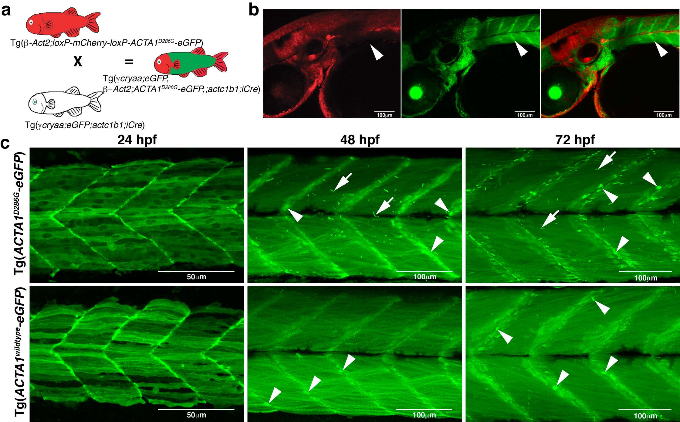
Fig. S1 Generation and phenotypic characterization of Tg(ACTA1-eGFP) stable lines. a) Crossing scheme to generate Tg(ACTA1-eGFP) fish. b) Confocal images show that upon iCre-mediated deletion of the loxP cassette, containing mCherry, eGFP is localized to the muscle (green) and does not overlap with other tissues (red; arrowheads). c) Maximum projection confocal microscopy images showing expression of ACTA1-eGFP in zebrafish skeletal muscle at 24, 48, and 72 hpf. Tg(ACTA1D286G-eGFP)high expression results in the formation of nemaline bodies (arrows) at 48 hpf and aggregates at the myosepta (arrowheads). No nemaline bodies are observed in Tg(ACTA1wildtypeeGFP)high skeletal muscle at any stage however globular aggregates are evident from 48 hpf at the myosepta (arrowheads).