
IMAGE
Fig. 6
- ID
- ZDB-IMAGE-150929-24
- Genes
- Publication
- Lippok et al., 2014 - Pou5f1 protein expression and posttranslational modification during early zebrafish development
- All Figures
- Figures for Lippok et al., 2014
Image
|
Figure Caption
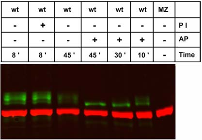
Fig. 6
Pou5f1 modification by phosphorylation. Western blot analysis of 70% epiboly stage embryo extracts treated at 37°C with phosphatase inhibitor (PI) or alkaline phosphatase (AP) for defined periods (indicated at bottom). Pou5f1-positive protein bands were detected around 60 kDa in wild-type (WT) but reduced to a band of approximately 57 kDa molecular mass upon AP treatment. Anti-α-tubulin was used as loading control (red, 55 kDa).
Figure Data
Acknowledgments
This image is the copyrighted work of the attributed author or publisher, and
ZFIN has permission only to display this image to its users.
Additional permissions should be obtained from the applicable author or publisher of the image.
Full text @ Dev. Dyn.