
Fig. 4
- ID
- ZDB-IMAGE-150929-22
- Genes
- Publication
- Lippok et al., 2014 - Pou5f1 protein expression and posttranslational modification during early zebrafish development
- All Figures
- Figures for Lippok et al., 2014
|
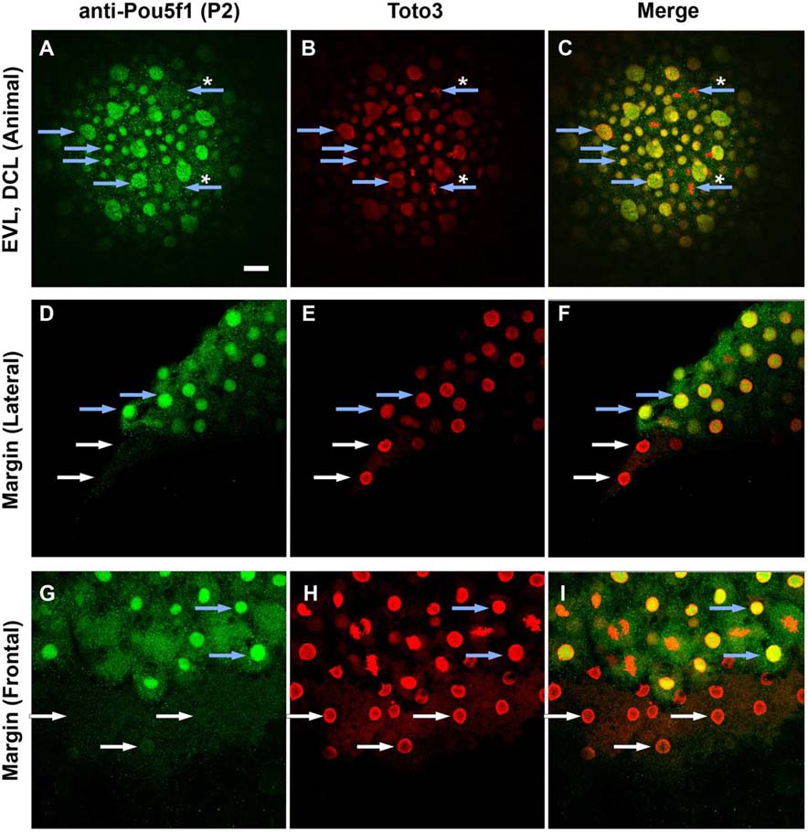
Fig. 4
Distribution of Pou5f1 in enveloping layer (EVL), deep cells and yolk syncytial layer (YSL) of wild-type (WT) embryos. A–I: Confocal images of anti-Pou5f1 immunofluorescence (green) WT whole-mount embryos at 90% epiboly (A–C) and sphere (D–I) stages in which all cell nuclei were stained with Toto3 (red). Blue arrows indicate blastoderm cell nuclei and white arrows indicate yolk syncytial nuclei. Large nuclei in A–C are EVL nuclei. Pou5f1 was not localized to chromatin in dividing blastomeres (blue arrows with asterisk), and also not in yolk syncytial nuclei (white arrows). Blastoderm layer at the animal pole (A–C), lateral margin (D–F), and frontal margin (G–I) are shown.