
IMAGE
Fig. 2
- ID
- ZDB-IMAGE-150929-20
- Genes
- Publication
- Lippok et al., 2014 - Pou5f1 protein expression and posttranslational modification during early zebrafish development
- All Figures
- Figures for Lippok et al., 2014
Image
|
Figure Caption
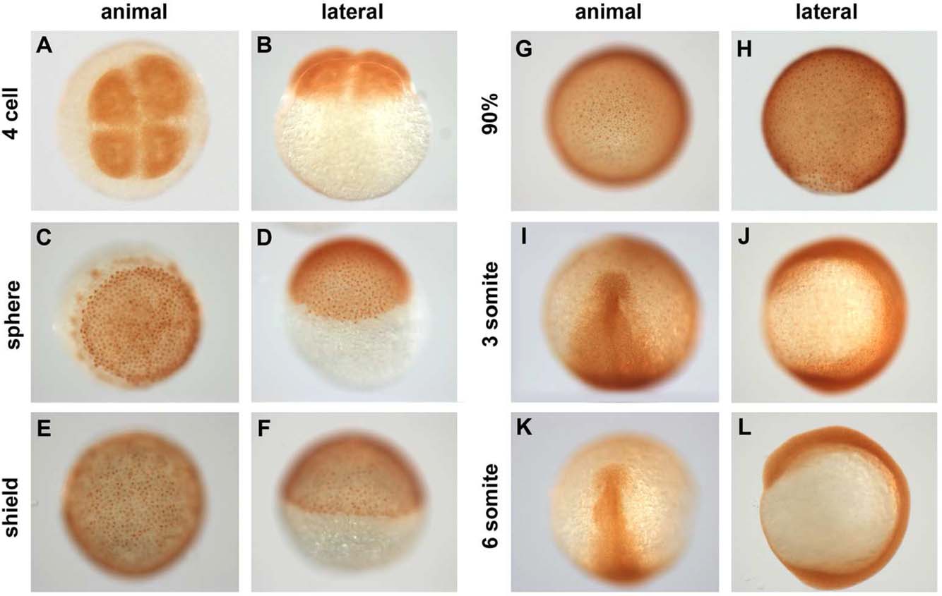
Fig. 2
Spatiotemporal distribution of Pou5f1 in wild-type (WT) embryos. A–L: Pou5f1 distribution in whole-mount WT embryos from four-cell to six-somite stages analyzed by anti Pou5f1 HRP/DAB immunohistochemistry. Animal views (A,C,E,G,I,K), dorsal to the bottom (E,G,I,K), and lateral views (B,D,F,H,J,L), dorsal to the right (F,H,J,L).
Figure Data
Acknowledgments
This image is the copyrighted work of the attributed author or publisher, and
ZFIN has permission only to display this image to its users.
Additional permissions should be obtained from the applicable author or publisher of the image.
Full text @ Dev. Dyn.