
|
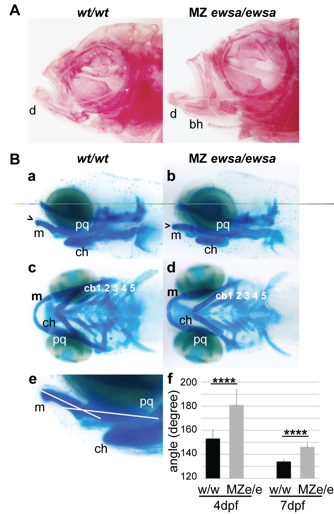
Fig. 2
4 dpf MZ ewsa/ewsa mutants display an aberrant angle of Meckel’s cartilage and palatoquadrate.
A. Lateral views (anterior to the left) of wt/wt (left) and MZ ewsa/ewsa (right) and ventral views of adult zebrafish. The calcified bones were visualized by alizarin red staining. B. Lateral views (anterior to the left) of (a) wt/wt and (b) MZ ewsa/ewsa and ventral views of (c) wt/wt and (d) MZ ewsa/ewsa chondrocytes from 4 dpf zebrafish embryos visualized with alcian blue. (e and f) Angle formed by Meckel’s cartilage showing that the palatoquadrate is wider in the MZ ewsa/ewsa mutant than wt/wt at 4 dpf and 7 dpf. bh: basihyal, d: dentary, m: Meckel’s cartilage, pq: palatoquadrate, ch: ceratohyal, cb: ceratobranchial.