
IMAGE
Fig. 4, S1
- ID
- ZDB-IMAGE-150430-45
- Publication
- Lush et al., 2014 - ErbB expressing Schwann cells control lateral line progenitor cells via non-cell-autonomous regulation of Wnt/beta-catenin
- All Figures
- Figures for Lush et al., 2014
Image
|
Figure Caption
Fig. 4, S1
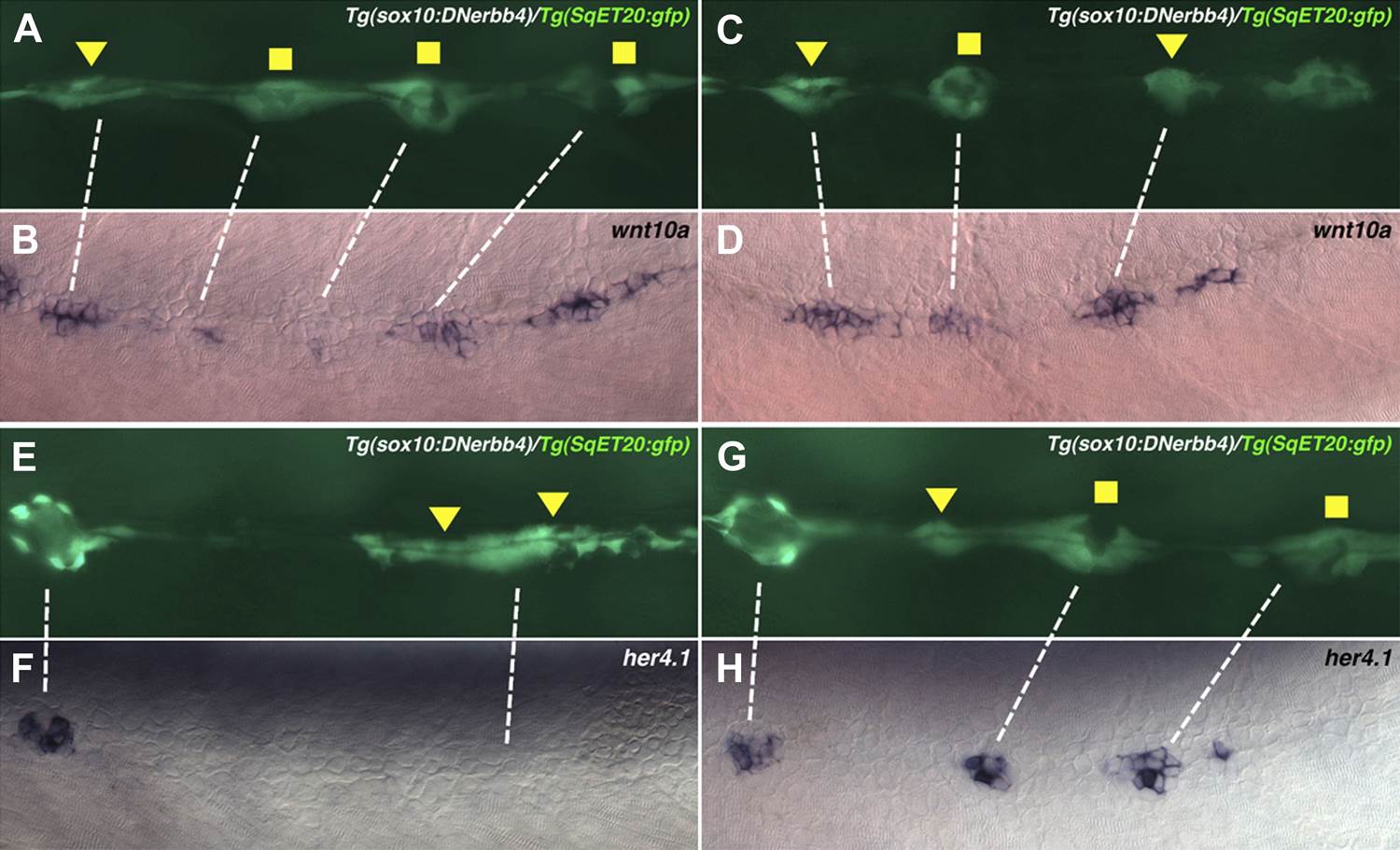
wnt10a is expressed in proliferating interneuromast cells while her4.1 is expressed in differentiating and mature neuromasts.
Lateral line cells from Tg(sox10:DNerbb4)/Tg(SqET20:gfp) were imaged at 48 hpf. Larvae were then fixed at processed for wnt10a or her4.1 in situ hybridization and photographed at the same level. (A–D) Proliferating interneuromast cells (arrowhead) express higher wnt10a than more mature intercalary neuromasts (square). (E–H) her4.1 is expressed in more mature intercalary neuromasts (squares), but is absent from more immature proliferating interneuromast cells (arrowhead).
Acknowledgments
This image is the copyrighted work of the attributed author or publisher, and
ZFIN has permission only to display this image to its users.
Additional permissions should be obtained from the applicable author or publisher of the image.
Full text @ Elife