
|
Fig. S2
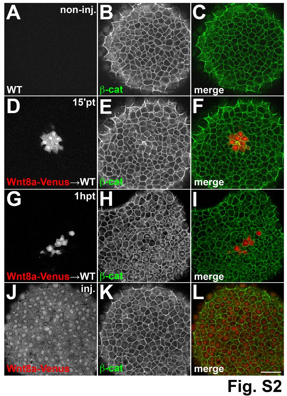
Time dependent β-catenin response to Wnt8a. Single confocal sections at the animal pole of shield stage embryos stained with an antibody against β-catenin. (A–C) β-catenin staining is restricted to the membrane in a WT embryo. (D–I) Cells derived from embryos injected with a lineage tracer (red) and wnt8a-venus mRNA transplanted into WT host embryos. In embryos fixed 15 minutes after transplantation nuclear β-catenin is not detected around the transplanted cells (D–E). In embryos fixed 1 hour after transplantation strong nuclear β-catenin is detected around transplanted cells (G–I). Embryos injected with a lineage tracer (red) and wnt8a-venus RNA at the one-cell stage have no nuclear β-catenin (J-L). (A,D) Red channel, (B,E) green channel, (C,F) overlay. Scale bars: 50 µm.