Fig. 1
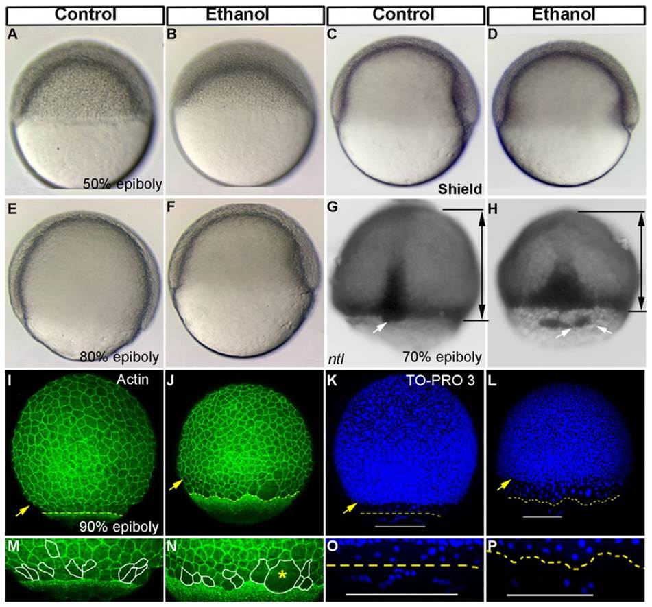
Ethanol exposure reduces epiboly progression and dorsal forerunner cell aggregation.
(A–F) Live embryos at 50% epiboly (A,B), shield (C,D), and 80% epiboly stages (E,F) showed reduced epiboly progression in the ethanol treated embryos (B,D,F) compared to control (A,C,E). (G,H) In situ hybridization depicting ntl showed epiboly delay in the deep cells and obvious separation of the dorsal forerunner cells from the deep cell margin in the ethanol treated embryo (H). Black lines with arrows indicate the distance between the deep cell margin and the animal pole. White arrows: dorsal forerunner cells. (I,J) 3D renderings of confocal microscopy optical sections of phalloidin stained (F-actin) gastrulae. Yellow arrowhead: deep cell margin; yellow perforated line: EVL margin. (K,L) 3D renderings of confocal microscopy optical sections of TO-PRO-3 stained embryos showed deep cells nuclei, EVL cell nuclei and YSL nuclei. Yellow arrowhead: deep cell margin; yellow perforated line: EVL margin drawn from F-actin staining (I,J); white line: yolk syncytial nuclei margin. (M,N) High magnification images of control and ethanol treated embryos highlighting cell boundaries of a few EVL cells. Cells at the embryo margins in the control embryo showed elongated EVL cells, roughly perpendicularly aligned to the EVL margin (M). Ethanol treated embryos showed rounder and not correctly aligned EVL cells (N). Yellow asterisk indicates big multinucleated cell. (O,P) High magnification images of the TO-PRO-3 stained control and ethanol treated embryos. Control embryos showed YSL nuclei proceeded beyond the EVL (O). Ethanol treated embryos showed fewer YSL nuclei proceeded beyond the EVL.

