
Image
|
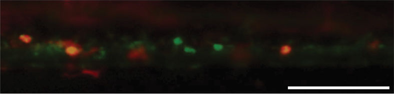
Figure Caption
Fig. 4
A549 cells labelled with DiO (green) were injected into the yolk sac of an fms:mCherry zebrafish at 48 h post-fertilization. Five days later partial overlap between DiO (A549 cells) and mCherry (macrophages) signals is detected (yellow), indicating that dye transfer has occurred, probably as a consequence of phagocytosis of dead A549 cells. Scale bar, 100 µm.
Acknowledgments
This image is the copyrighted work of the attributed author or publisher, and
ZFIN has permission only to display this image to its users.
Additional permissions should be obtained from the applicable author or publisher of the image.
Full text @ Interface Focus