
|
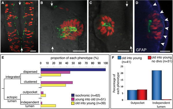
Fig. 5
Heterochronic transplanted cells integrate into the host neuroepithelium, and can generate ectopic lumen surfaces. (A-D) Dorsal view projected confocal z-series of transplanted cells in the host hindbrain at 24 hours post fertilization (hpf). White arrows indicate the midline of neural tube. Scale bar is 25 µm. (A) By 24 hpf, isochronic transplanted cells have established apicobasal polarity, as revealed by green fluorescent protein/polarity protein partitioning defective 3 fusion (Pard3-GFP) localization at the ventricular surface of the neuroepithelium. (B) Heterochronic transplanted cells can generate ectopic apical surfaces that form an outpocket of the host neuroepithelium. (C) Heterochronic transplanted cells can form rosettes with a lumen independent of the host lumen. (D) Glial fibrillary acidic protein (GFAP) (blue) staining confirms that an ectopic rosette of transplanted cells is located within the neuroepithelium. Arrowheads show lateral edge of neural tube. (E) Frequency histogram showing the proportion of isochronic and heterochronic transplanted cells that fall into the four phenotypes of integrated dispersed, integrated clustered, ectopic lumen outpocket or independent lumen. (F) Graph showing that blocking cell division abolishes formation of independent lumens in old into young transplants.