
Fig. 1
- ID
- ZDB-IMAGE-150422-28
- Publication
- Mackay et al., 2015 - Vitamin K reduces hypermineralisation in zebrafish models of PXE and GACI
- All Figures
- Figures for Mackay et al., 2015
|
Fig. 1
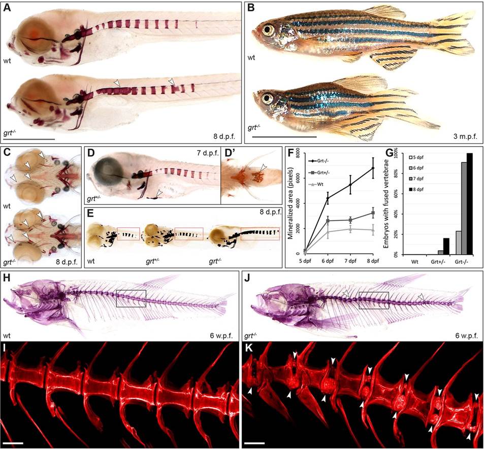
The gräte mutant phenotype is characterised by hypermineralisation in the skin and axial skeleton. (A) Alizarin Red staining of embryos at 8days post-fertilisation (dpf), demonstrating hypermineralisation along the vertebral column (arrowheads). Scale bar: 1mm. (B) Adult fish are viable, but feature a curved spine and reduced length. Scale bar: 1cm. (C) grt-/- embryos (ventral view) with enhanced mineralisation in craniofacial elements (arrowheads). (D) Skin mineralisation is infrequently seen in grt+/- and grt-/- embryos (D′, ventral view). (E) Representative images showing the area quantified by the mineralisation assay used in this and in subsequent figures. (F) Vertebral mineralisation in grt-/- embryos proceeds faster than in wild type, leading to (G) vertebral fusion from 6dpf onwards. (H,J) Alizarin Red staining at 6weeks post fertilisation (wpf) reveals a thickened, curved spine in grt-/- fish; confocal images (I,K) of the boxed regions reveal mineralised nodules on the margins of the intervertebral space (arrowheads). Scale bars: 0.1mm.