
Image
|
Figure Caption
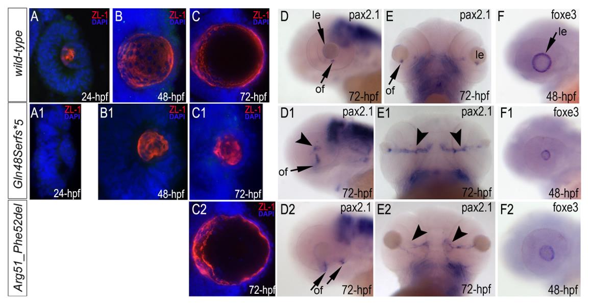
Fig. S5
Immunostaining with ZL-1 (red) and in situ hybridization with pax6b, pax2.1 and foxe3 antisense riboprobes in wild-type (A-F), mab21l2Q48Sfs*5 (A1-F1) andmab21l2R51_F52del (C2-F2) embryos.
The ZL-1 staining is absent in 24-hpf (A1) but present in 48–72-hpf mutant embryos (B1, C1, C2), pax2.1 pattern is abnormal in 72-hpf mutant embryos (D1, E1, D2, E2), arrowhead in D1 shows abnormal areas of pax2.1-positive cells in the central retina and arrowheads in E1 show broad and intense expression in the region of optic fissure; foxe3 expression is decreased in 48-hpf mutants (F1, F2); le, lens; of, optic fissure.
Figure Data
Acknowledgments
This image is the copyrighted work of the attributed author or publisher, and
ZFIN has permission only to display this image to its users.
Additional permissions should be obtained from the applicable author or publisher of the image.
Full text @ PLoS Genet.