
|
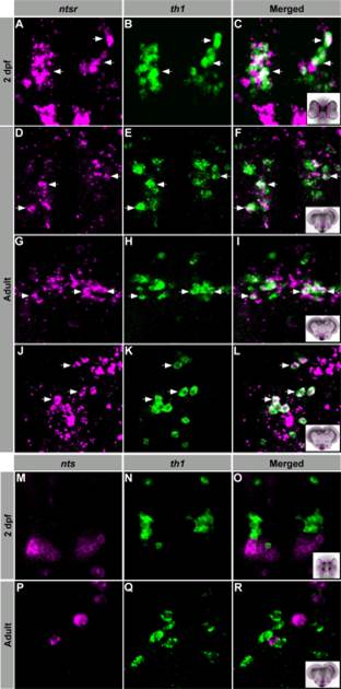
Fig. 4
Dopaminergic neurons express NTSR. Confocal imaging of double fluorescent ISH experiments in larvae and adult zebrafish. A–L: 2 dpf larvae and transversal adult brain sections showing colocalization of ntsr- (magenta) and th1- (green) expressing cell bodies within the TP and H (A–C), TPp (D–F), Hd (G–I), and Hc (J–L). M–R: 2 dpf larvae (M–O) and transversal adult brain sections (P–R) showing nts- (magenta) and th1- (green) expressing cell bodies within the hypothalamus. Arrows indicate representative coexpressing cells (white). All images are single confocal sections. TPp, periventricular nucleus of the posterior tuberculum; Hd, dorsal zone of the periventricular hypothalamus; Hc, caudal zone of the periventricular hypothalamus.