
Fig. 3
|
Fig. 3
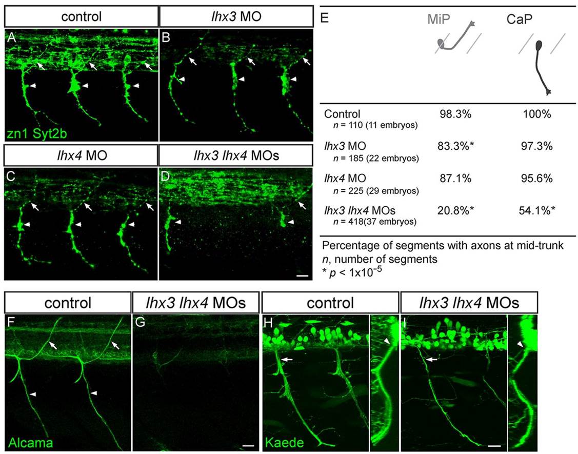
Lhx3 and Lhx4 are required for normal PMN axon morphology. (A-D) Lateral views of 28hpf embryos co-labeled with zn1 and anti-Syt2b. Control (A), lhx3 MO-injected (B) and lhx4 MO-injected (C) embryos have normal dorsal MiP axons (arrows) and ventral CaP axons (arrowheads). MiP, CaP, or MiP and CaP axons are often absent from lhx3+lhx4 MO-injected embryos (D). Apparent variation in intraspinal labeling in B and C was a consequence of embryo orientation and does not reflect differences in IN axon labeling. (E) Quantification of MiP and CaP phenotypes in control and MO-injected embryos. (F,G) Lateral views of somites 8-12 of 72hpf embryos labeled with anti-Alcama. Alcama labeling of SMN axons and somata was almost completely missing from embryos injected with lhx3+lhx4 MOs. (H,I) Lateral views of 48hpf s1020t;Tg(UAS-E1b:Kaede) embryos. Ventral nerves in lhx3+lhx4 MO-injected embryos were thinner (arrows), yet exited through the ventral root (arrowhead); inset shows an optical cross-section. Scale bars: 20µm.