
Image
|
Figure Caption
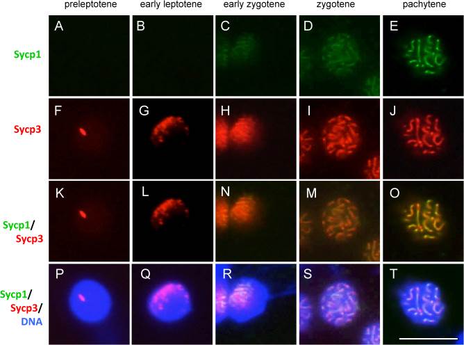
Fig. 5
Immunocytochemical analyses of Sycp1 and Sycp3 in primary spermatocytes. Localization of Sycp1 (A–E), Sycp3 (F–J), merged Sycp1 and Sycp3 staining (K–O), and merged Sycp1, Sycp3, and nuclear staining with TO-PRO-3 (P–T) are shown at the preleptotene (A,F,K,P), early leptotene (B,G,L,Q), early zygotene (C,H,N,R), zygotene (D,I,M,S), and pachytene (E,J,O,T) stages. Each developmental stage of spermatocytes was identified according to their synaptonemal complex (SC) structures. Bar = 10 µm.
Figure Data
Acknowledgments
This image is the copyrighted work of the attributed author or publisher, and
ZFIN has permission only to display this image to its users.
Additional permissions should be obtained from the applicable author or publisher of the image.
Full text @ Dev. Dyn.