
|
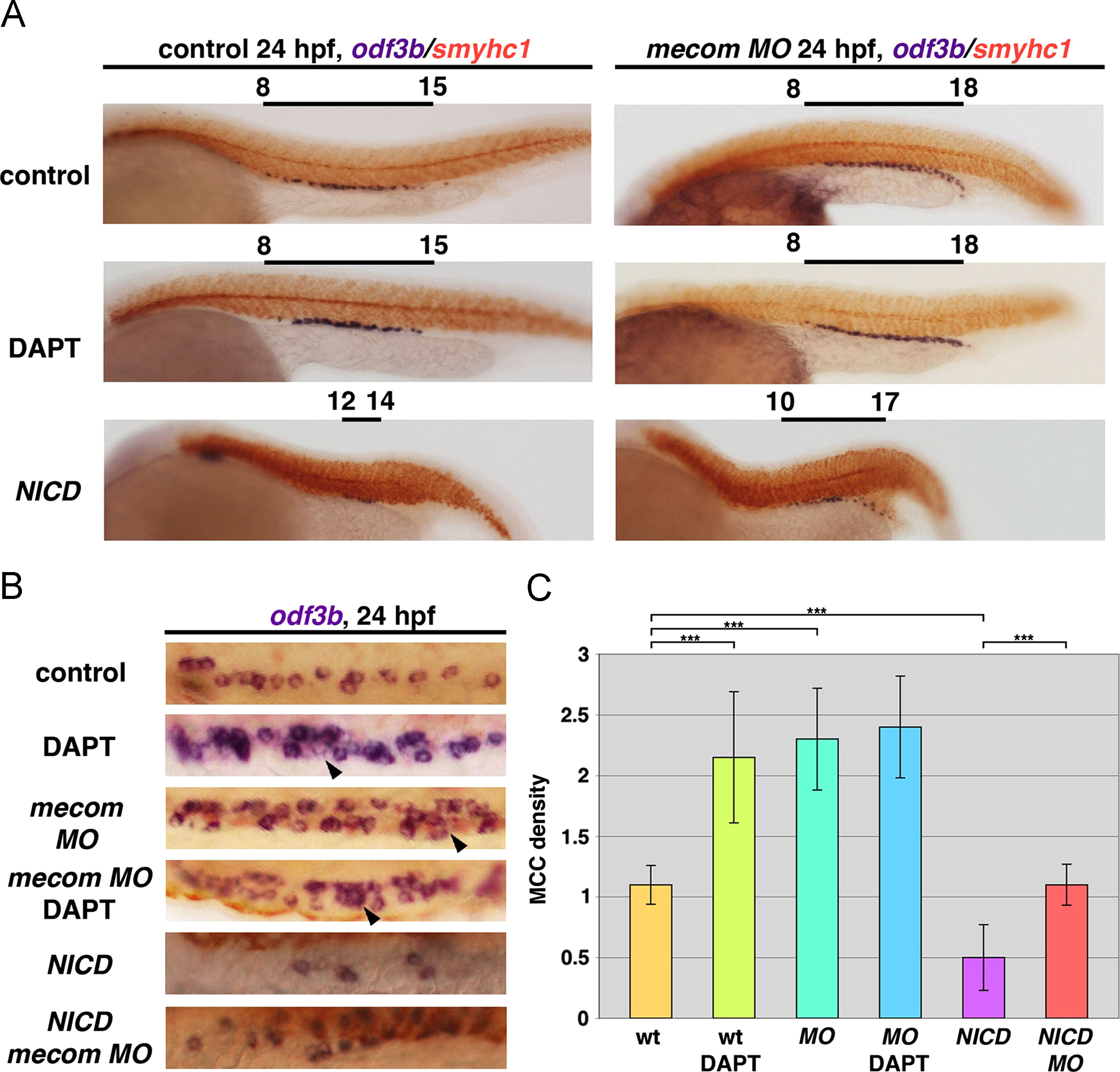
Fig. 7
mecom acts upstream of Notch signaling to modulate MCC differentiation and regulate the MCC domain. (A) Wild type embryos treated with 100 µM DAPT showed a significant increase of MCC number without ectopic MCC formation. mecom knockdown led to a caudal expansion of MCC in the DL and PD, and exhibited increased MCC density compared to wild types. A similar condensed MCC arrangement could also be seen in mecom morphants treated with DAPT. The overexpression of Notch1a resulted in decreased MCCs in heatshock induced Tg(hsp70:gal4; uas:notch1a-intra) embryos. Ectopic MCC formation associated with mecom knockdown was abolished by Notch signaling activation, though the domain of MCCs was still expanded. (B) Differentiated MCCs at 24 hpf under 10× magnification in a single nephron from wild types, wild types treated with DAPT, mecom morphants and morphants treated with DAPT, and finally wild types and mecom morphants with NICD overexpression. Note: MCCs displayed a condensed organization/cluster pattern in DAPT-treated wild type and mecom morphant pronephros, while DAPT treatment in mecom morphants failed to induce further MCC density. Arrows indicate large MCC aggregates observed in DAPT-treated wild types, mecom morphants, and DAPT-treated mecom morphants, which were absent from the wild type pronephros. For each experiment, at least 20 embryos were examined. (C) Quantification of MCC density in wild types, wild type embryos treated with DAPT, mecom morphants, and mecom morphants treated with DAPT. The Student t-test revealed significant increase of MCC density in DAPT treated wild types and mecom morphants relative to untreated wild types (***p=0.0005). Alterations of MCC density between mecom morphants and morphants treated with DAPT did not reach statistical significance. For each experiment, at least 20 embryos were examined.
Reprinted from Developmental Biology, 386(1), Li, Y., Cheng, C.N., Verdun, V.A., and Wingert, R.A., Zebrafish nephrogenesis is regulated by interactions between retinoic acid, mecom, and Notch signaling, 111-122, Copyright (2014) with permission from Elsevier. Full text @ Dev. Biol.