
Fig. 5
- ID
- ZDB-IMAGE-150325-73
- Publication
- Verleyen et al., 2014 - Orphan G-Protein Coupled Receptor 22 (Gpr22) Regulates Cilia Length and Structure in the Zebrafish Kupffer's Vesicle
- All Figures
- Figures for Verleyen et al., 2014
|
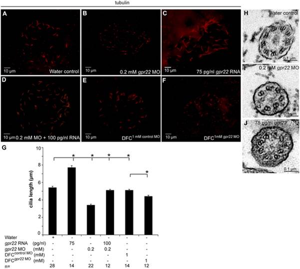
Fig. 5
Gpr22 affects cilia length and structure in the KV.
(A–F) Immunostaining with anti-acetylated tubulin antibody on embryos at 10 somite stage injected at (A–D) one cell stage with (A) water, (B) 0.2 mM gpr22 MO, (C) 75 pg/nl gpr22 RNA, (D) 0.2 mM gpr22 MO and 100 pg/nl gpr22 RNA, or injected at (E, F) mid-blastula stage with (E) 1 mM control MO, (F) 1 mM gpr22 MO. Shown are the cilia of the KV. (G) Quantification of average cilia length ± s.d. (B, G) Whole embryo or (F, G) DFC-specific knock down of gpr22 significantly reduces cilia length. (C, G) In contrast, gpr22 overexpression results in significantly longer cilia. (D, G) Co-injection of 0.2 mM MO and 100 pg/nl RNA almost completely rescues cilia length. (H–J) TEM of cross sections of the KV cilia at 10 somite stage. (H) Structure of water control KV cilia showing 9 parallel outer microtubule doublets. (I) gpr22 knock down or (J) overexpression results in a disruption of the proper microtubuli arrangement or (I) even an absence of doublets. MO = morpholino, n = number of analyzed embryos, DFC = dorsal forerunner cells, DFCsuperscript = injected at mid-blastula stage, KV = Kupffer’s vesicle, TEM = transmission electron microscopy, * = P<0.01 (unpaired, two-tailed T-test).