
Fig. 2
- ID
- ZDB-IMAGE-150325-47
- Genes
- Publication
- Kenyon et al., 2015 - Zebrafish Rab5 Proteins and a role for Rab5ab in nodal signalling
- All Figures
- Figures for Kenyon et al., 2015
|
Fig. 2
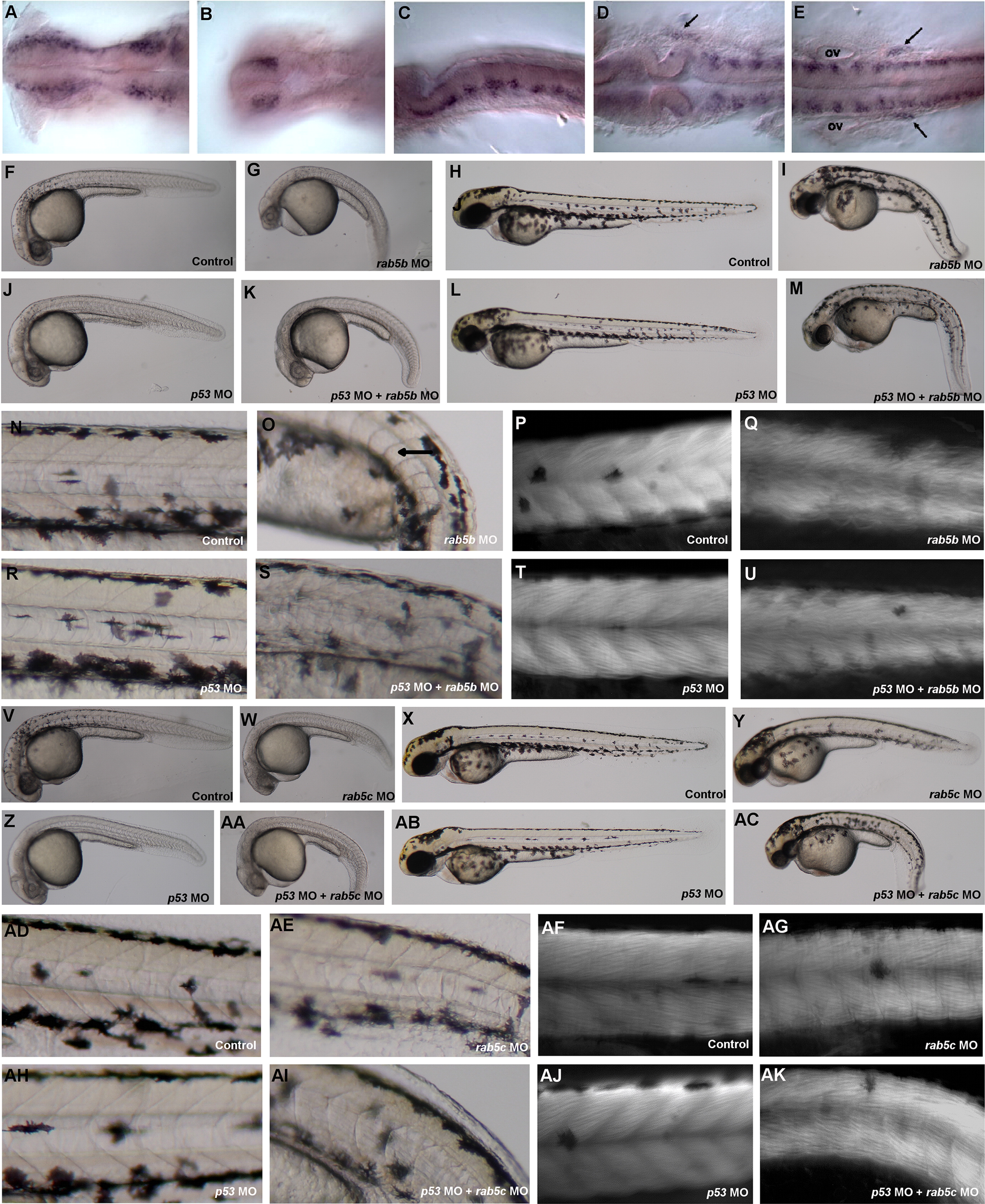
Expression and loss of function of the rab5a family. (A) Expression of rab5aa in the forebrain and midbrain region of a 24 hpf embryo. (B) Forebrain region with dorsal focus showing two patches of bilateral telencephalic cells. (C) Hindbrain region showing expression on the central region of each rhombomere. (D) Expression of rab5aa in cells outside the neural tube at the level of the midbrain/hindbrain boundary (arrow). (E) Expression of rab5aa at the end of the hindbrain (arrows) and in the trunk of the embryo. (F) Side view of a 24 hpf embryo injected with 10 ng of control MO (n=205/207). (G) Side view of a 24 hpf embryo injected with 8 ng of rab5b MO (n=141/143). (H) Side view of a 48 hpf embryo injected with 10 ng of control MO (n=204/204). (I) Side view of a 48 hpf embryo injected with 8 ng of rab5b MO (n=117/126). (J) Side view of a 24 hpf embryo injected with 12 ng of p53 MO (n=77/85). (K) Side view of a 24 hpf embryo co-injected with 12 ng of p53 MO and 8 ng of rab5b MO (n=98/108) (L) Side view of a 48 hpf embryo injected with 12 ng of p53 MO. (M) Side view of a 48 hpf embryo co-injected with 12 ng of p53 MO and 8 ng of rab5b MO (n=52/62). (N) Magnification of trunk region showing somites and notochord in 48 hpf control-injected embryos. (O) Magnification of trunk region showing somites and notochord in 48 hpf rab5b MO-injected embryos. (P) Magnification of trunk region showing somites in a 48 hpf control-injected embryo stained with phalloidin. (Q) Magnification of trunk region showing somites in a 48 hpf rab5b MO-injected embryo stained with phalloidin. (R) Magnification of trunk region showing somites and notochord in 48 hpf p53 MO injected embryos. (S) Magnification of trunk region showing somites and notochord in 48 hpf p53 MO and rab5b MO co-injected embryos. (T) Magnification of trunk region showing somites in a 48 hpf p53 MO injected embryo stained with phalloidin. (U) Magnification of trunk region showing somites in a 48 hpf p53 MO and rab5b MO co-injected embryo stained with phalloidin. (V) Lateral view of a 30 hpf embryo injected with 5 ng of control MO (n=92/95). (W) Lateral view of a 30 hpf embryo injected with 6 ng of rab5c MO (n=174/175). (X) Lateral view of a 48 hpf embryo injected with 5 ng of control MO (n=92/95). (Y) Lateral view of a 48 hpf embryo injected with 6 ng rab5c MO (n=158/159). (Z) Lateral view of a 30 hpf embryo injected with 9 ng of p53 MO (n=54/54). (AA) Lateral view of a 30 hpf embryo co-injected with 9 ng of p53 MO and 6 ng of rab5c MO (n=n=54/56). (AB) Lateral view of a 48 hpf embryo injected with 9 ng of p53 MO. (AC) Lateral view of a 48 hpf co-injected with 9 ng of p53 MO and 6 ng of rab5c MO (n=37/48). (AD) Magnification of trunk region showing somites and notochord in 48 hpf control-injected embryos. (AE) Magnification of trunk region showing somites and notochord in 48 hpf rab5c MO-injected embryos. (AF) Magnification of trunk region showing somites in a 48 hpf control-injected embryo stained with phalloidin. (AG) Magnification of trunk region showing somites in a 48 hpf rab5c MO-injected embryo stained with phalloidin. (AH) Magnification of trunk region showing somites and notochord in 48 hpf p53 MO injected embryos. (AI) Magnification of trunk region showing somites and notochord in 48 hpf p53 MO and rab5c MO co-injected embryos. (AJ) Magnification of trunk region showing somites in a 48 hpf p53 MO injected embryo stained with phalloidin. (AK) Magnification of trunk region showing somites in a 48 hpf p53 MO and rab5c MO co-injected embryo stained with phalloidin. (A, B, D, E are dorsal views, anterior to the left and the eyes were manually removed for simplification C is a side view, anterior to the left (‘ov’ indicates otic vesicle).
Reprinted from Developmental Biology, 397(2), Kenyon, E.J., Campos, I., Bull, J.C., Williams, P.H., Stemple, D.L., Clark, M.D., Zebrafish Rab5 Proteins and a role for Rab5ab in nodal signalling, 212-24, Copyright (2015) with permission from Elsevier. Full text @ Dev. Biol.