
Fig. S7
- ID
- ZDB-IMAGE-150318-21
- Publication
- Gerlach et al., 2014 - Zebrafish pronephros tubulogenesis and epithelial identity maintenance are reliant on the polarity proteins Prkc iota and zeta
- All Figures
- Figures for Gerlach et al., 2014
|
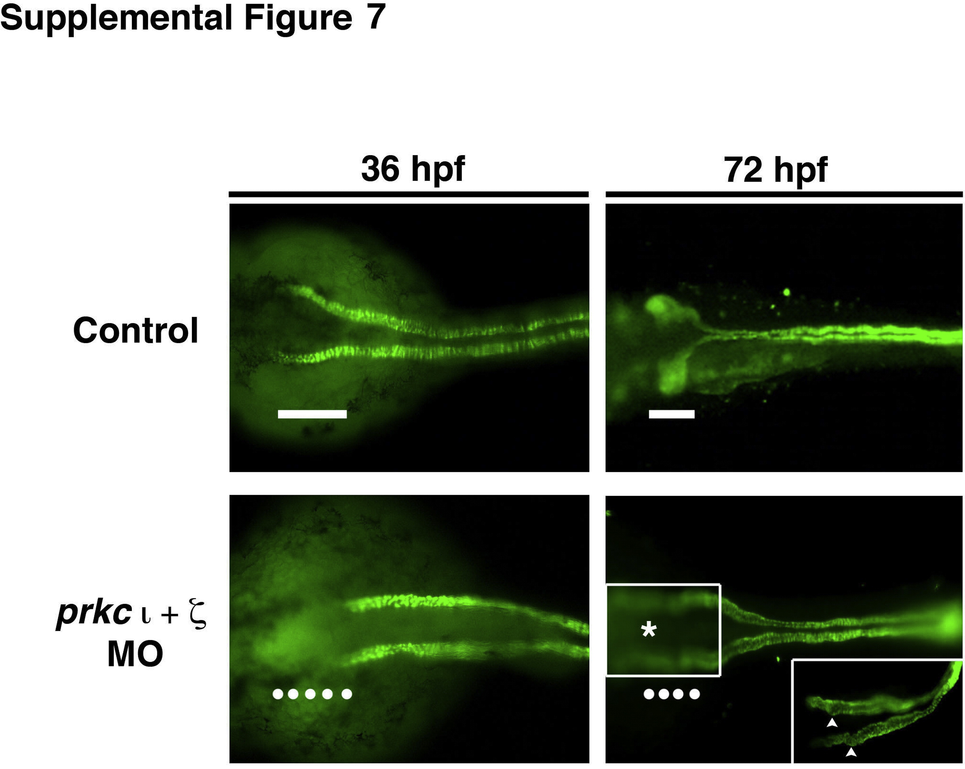
Fig. S7 prkCI/ζ deficient embryos display reduced protein expression of the Na+/K+ ATPase in the neck region of the pronephros. Based on whole mount IF, wild-type embryos at 36 hpf exhibited Na+/K+ ATPase protein expression along the entire length of the pronephric tubule, including the proximal-most region (white bar). In contrast, double prkCI/ζ morphants at the 36 hpf stage fail to show Na+/K+ ATPase in the proximal most region of the pronephros (line of white dots). In wild-type embryos at 72 hpf, Na+/K+ ATPase continues to demarcate the entire tubule and shows the convoluted nature of the PCT (white bar), while prkCI/ζ deficient embryos have linear tubules that have failed to undergo normal morphogenesis. (Inset) Regions of the PCT in prkCI/ζ morphants exhibit folds and bulges (white arrowheads).
Reprinted from Developmental Biology, 396(2), Gerlach, G.F., Wingert, R.A., Zebrafish pronephros tubulogenesis and epithelial identity maintenance are reliant on the polarity proteins Prkc iota and zeta, 183-200, Copyright (2014) with permission from Elsevier. Full text @ Dev. Biol.