
Fig. 6
- ID
- ZDB-IMAGE-150318-10
- Publication
- Gerlach et al., 2014 - Zebrafish pronephros tubulogenesis and epithelial identity maintenance are reliant on the polarity proteins Prkc iota and zeta
- All Figures
- Figures for Gerlach et al., 2014
|
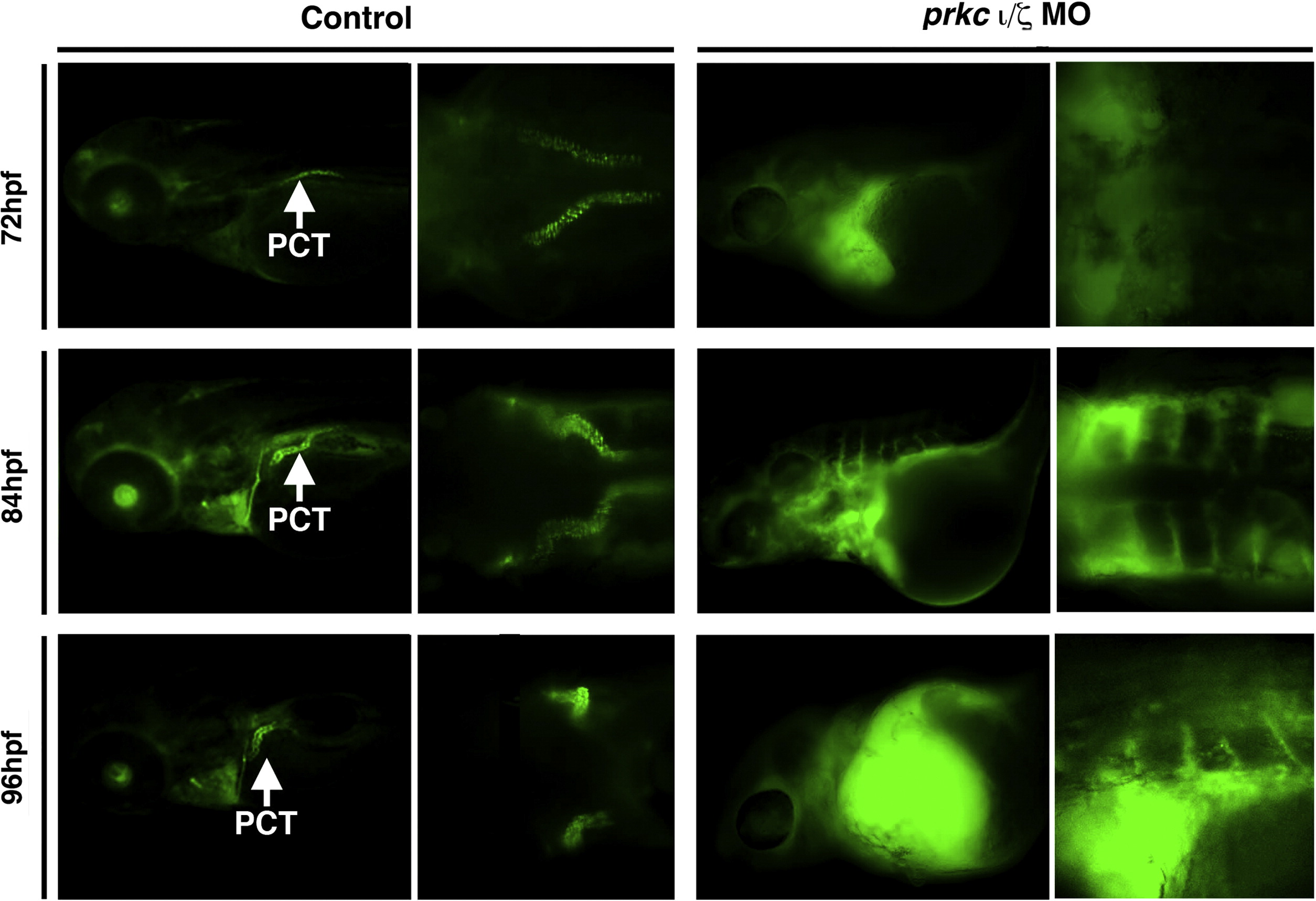
Fig. 6 Renal clearance and PCT endocytosis are abrogated in prkCI/ζ deficient embryos. Embryos were injected with 40-kDa dextran–FITC at 48 hpf, and imaged at 72, 84, and 96 hpf. Control wild-type embryos displayed renal clearance by diminution of the net fluorescent signal intensity over time, as well as dextran–FITC internalization throughout the PCT (white arrows) during its progressive morphogenesis. DoubleprkCI/ζ morphants displayed severe fluid accumulation, notably pericardial edema that remained strongly positive for the dextran–FITC conjugate label, and the PCT was not labeled by dextran endocytosis (left panels, lateral views; right panels, dorsal views, with exception of bottom right which shows a lateral/dorsal angled view).
Reprinted from Developmental Biology, 396(2), Gerlach, G.F., Wingert, R.A., Zebrafish pronephros tubulogenesis and epithelial identity maintenance are reliant on the polarity proteins Prkc iota and zeta, 183-200, Copyright (2014) with permission from Elsevier. Full text @ Dev. Biol.