
|
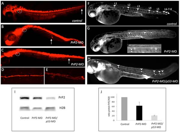
Fig. 1
PrP2 decrease expression results in abnormal PLL development.
A. In control embryos at 48 hours post fertilization (hpf) from nbt-dsred line that labels neurons and axons, the PLL nerve develops from the PLL ganglia until the tip of the tail (arrow). B–C. In PrP2-MO embryos, a range of defects for the PLL nerve is observed with premature arrest (B, C). D. Control PLL nerve fibers are tightly while PrP2-MO fibers exhibit abnormal branches were observed in severe cases (E). F. In control claudinB-GFP embryos at 48 hpf all derivatives issuing from the primordium express GFP and appear normal. Five regularly spaced neuromasts are present, with 3 terminal neuromasts at the tip of the tail. Due to embryo transparency, the neuromasts on the other side of the embryo are also visible. In PrP2-MO (G) and PrP2-MO/p53-MO (H) injected embryos (in order to avoid off-target defects), neuromast numbers are reduced, and often irregularly spaced. I–J. Western blot analysis of PrP2 expression using SAF84 antibody shows a decrease of the PrP2 protein after morpholino injections (n = 3 independent experiments).