
|
Fig. S5
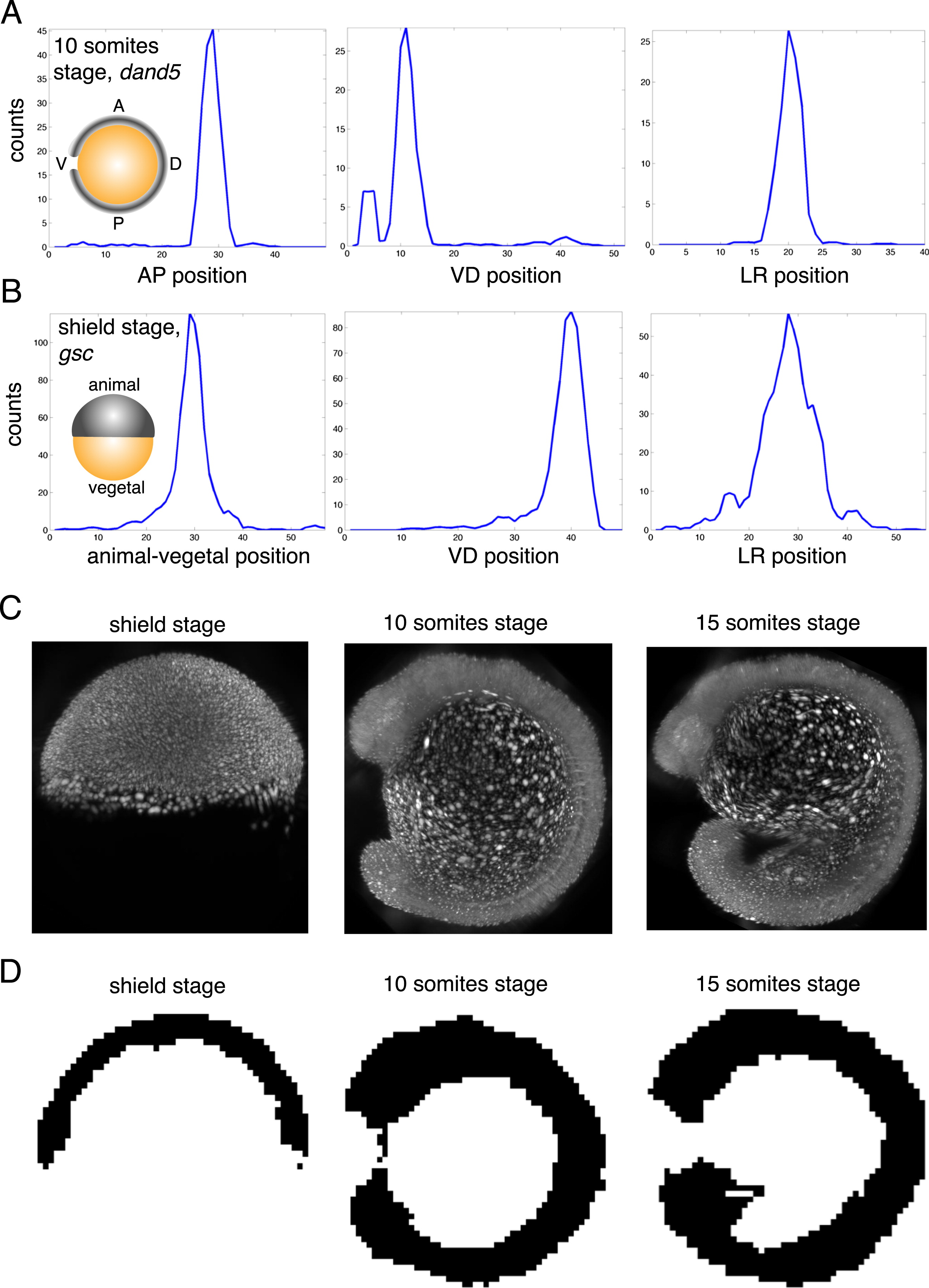
Selective Plane Illumination Microscopy Allows Measuring the Shape of Embryos in 3D, Related to >Figure 5
(A) Expression patterns for dand5 at 10 somites stage along AP, VD and LR axis.
(B) Expression patterns for gsc at shield stage along animal-vegetal, VD and LR axis.
(C) SPIM data for Tg(h2afva:h2afva-mCherry) zebrafish embryos at shield stage, 10 somites stage, and 15 somites stage. Images show projections along LR axis.
(D) 3D mask of zebrafish embryo at 3 stages (binary images created by aligning SPIM data for 3 different embryos). Images show single plane perpendicular to LR axis through the middle of the embryo.
Reprinted from Cell, 159, Junker, J.P., Noël, E.S., Guryev, V., Peterson, K.A., Shah, G., Huisken, J., McMahon, A.P., Berezikov, E., Bakkers, J., van Oudenaarden, A., Genome-wide RNA Tomography in the Zebrafish Embryo, 662-75, Copyright (2014) with permission from Elsevier. Full text @ Cell