IMAGE

Fig. 1

ID
ZDB-IMAGE-150123-5
Genes
Antibodies
Source
Figures for Du et al., 2012
Image
Figure Caption

Fig. 1 eomesafh105 mutant allele. Schematic of the 661 amino acid full-length Eomesa protein with T-domain in blue. Location of the stop codon in allele fh105 marked by asterisk. (B) Images of heterozygous (top) and homozygous (bottom) eomesafh105 adult fish. (C) Control western blot for the anti-Eomesa antibody, lanes as indicated. (D)Western blot of sphere stage wild type and MZeomesa embryos, lanes as indicated. 15 embryo equivalents loaded per lane. (E) Whole-mount in situ hybridization for eomesa on sphere stage embryos of the indicated genotypes.

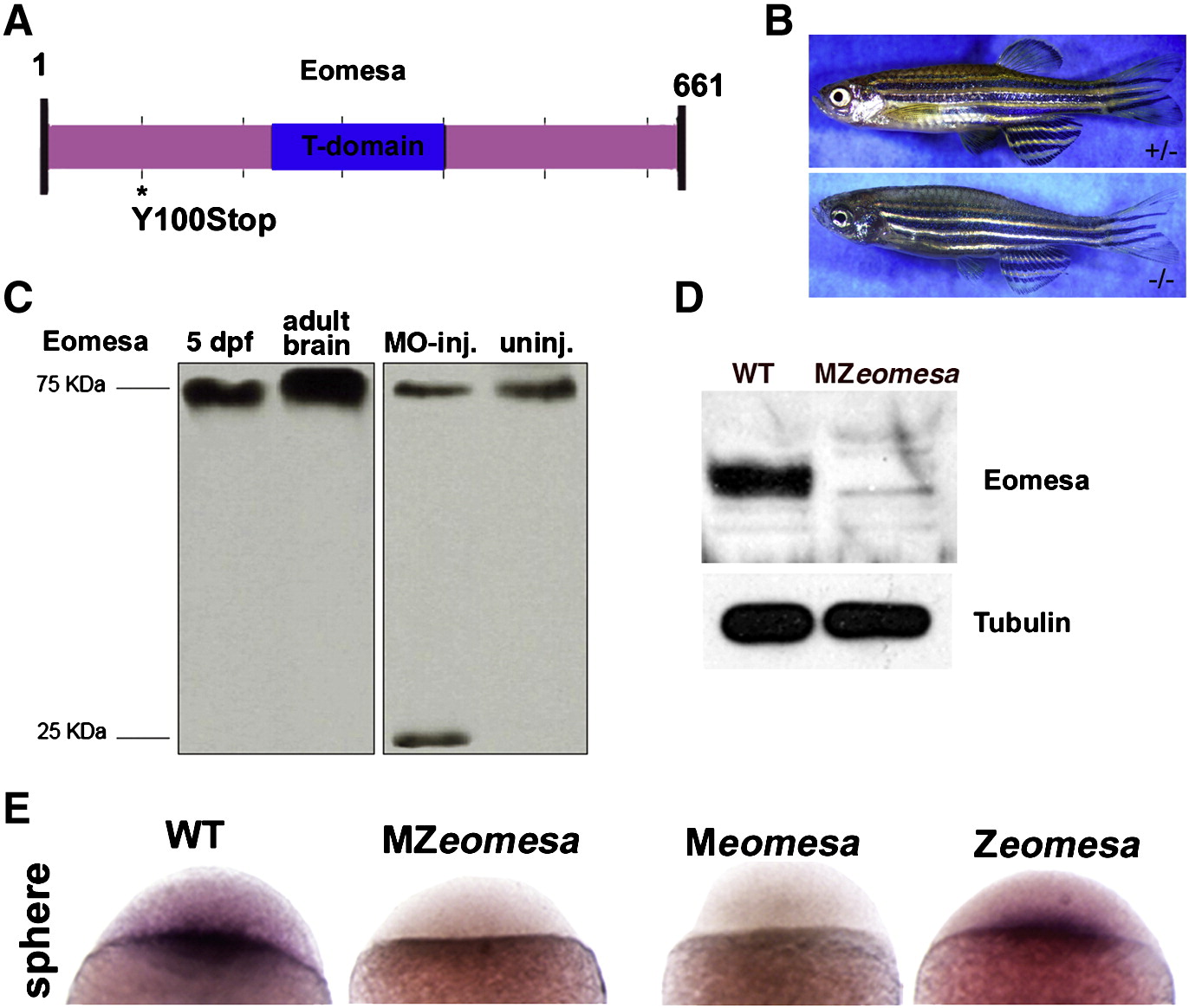
Figure Data
Acknowledgments
This image is the copyrighted work of the attributed author or publisher, and ZFIN has permission only to display this image to its users. Additional permissions should be obtained from the applicable author or publisher of the image.

Reprinted from Developmental Biology, 362(1), Du, S., Draper, B.W., Mione, M., Moens, C.B., and Bruce, A.E., Differential regulation of epiboly initiation and progression by zebrafish Eomesodermin A, 11-23, Copyright (2012) with permission from Elsevier. Full text @ Dev. Biol.