
Fig. 3
|
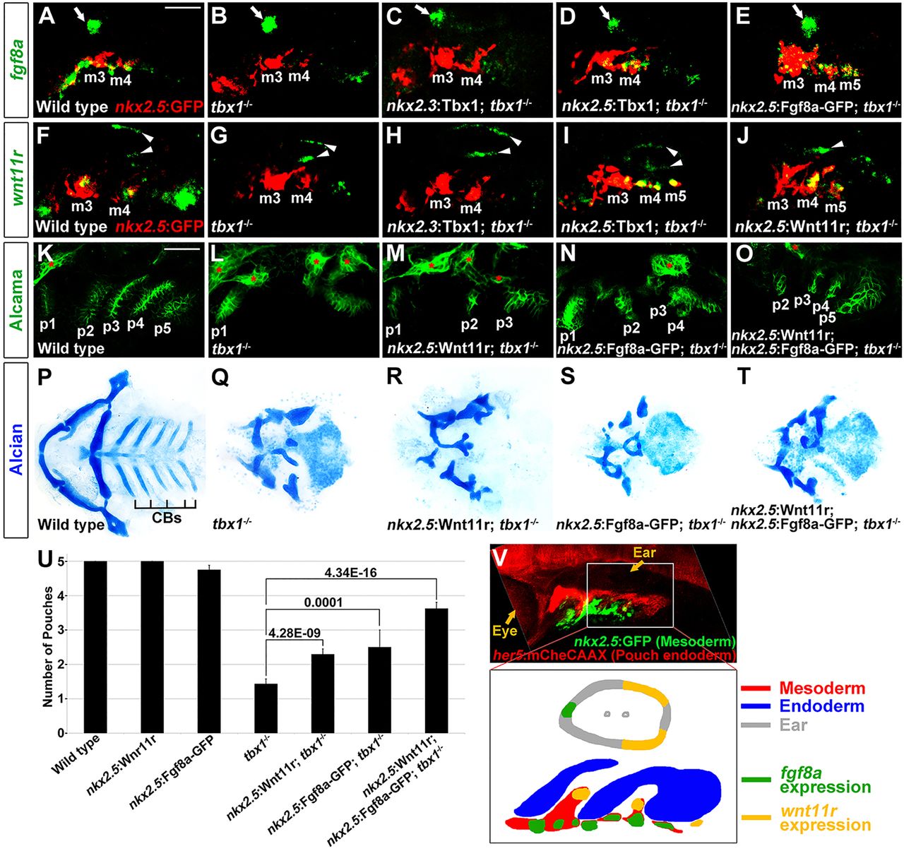
Fig. 3
Rescue of tbx1-/-pouch defects with mesodermal Fgf8a and Wnt11r. (A-J) Fluorescent in situ hybridization for fgf8a or wnt11r (green) and GFP immunohistochemistry to detect nkx2.5:GFP-positive mesoderm (red) at 30hpf. In wild type, fgf8a was expressed in ventral nkx2.5:GFP-positive mesodermal cores of arches 3 and 4 (m3 and m4), whereas wnt11r was in more dorsal subsets of these mesodermal cores. In tbx1 mutants, mesodermal expression of fgf8a (n=12 of 12) and wnt11r (n=10 of 11) was lost, although fgf8a and wnt11r expression in the anterior (arrow) and posterior (arrowheads) portions of the otic vesicle was unaffected. An nkx2.3:Tbx1 transgene did not restore mesodermal fgf8a (n=0 of 7) and wnt11r expression (n=0 of 5), whereas an nkx2.5:Tbx1 transgene restored mesodermal fgf8a (n=5 of 6) and wnt11r expression (n=9 of 9). Also, nkx2.5:Fgf8-GFP and nkx2.5:Wnt11r transgenes restored fgf8a (n=27 of 27) and wnt11r (n=21 of 21) expression, respectively. (K-O) Alcama immunohistochemistry (green) showed five pouches (p1-p5) in wild-type fish at 34hpf. tbx1-/- mutants lost all pouches except for the first (p1), whereas individual nkx2.5:Wnt11r or nkx2.5:Fgf8a-GFP transgenes modestly rescued, and combined nkx2.5:Wnt11r and nkx2.5:Fgf8a-GFP transgenes strongly rescued posterior pouches (p2-p5). Sensory ganglia are indicated with red asterisks. (P-T) Ventral views of dissected facial cartilages. A bilateral set of five CBs formed in wild-type zebrafish, and no CBs formed in tbx1 mutants. No rescue of CB cartilage was seen in nkx2.5:Wnt11r; tbx1-/- (n=39), nkx2.5:Fgf8a-GFP; tbx1-/- (n=34), or nkx2.5:Wnt11r; nkx2.5:Fgf8a-GFP; tbx1-/- larvae (n=31). (U) Quantification of pouch defects based on Alcama staining in wild type (n=51), nkx2.5:Wnt11r (n=49), nkx2.5:Fgf8a-GFP (n=36), tbx1-/- (n=62), nkx2.5:Wnt11r; tbx1-/- (n=44), nkx2.5:Fgf8a-GFP; tbx1-/- (n=37), and nkx2.5:Wnt11r; nkx2.5:Fgf8a-GFP; tbx1-/- (n=24). Data represent mean±s.e.m., P values are shown. (V) Low magnification view of an embryo at 32hpf showing nkx2.5:GFP-positive mesoderm (green) relative to her5:mCherryCAAX-positive pouch endoderm (red) and the developing eye and ear. The schematic shows expression of fgf8a (green) and wnt11r (yellow) within distinct subsets of nkx2.5-positive mesoderm (red) during the formation of endodermal pouches (blue). Scale bars: 40μm (A-O).