
Fig. 2
- ID
- ZDB-IMAGE-141125-2
- Publication
- Choksi et al., 2014 - Systematic discovery of novel ciliary genes through functional genomics in the zebrafish
- All Figures
- Figures for Choksi et al., 2014
|
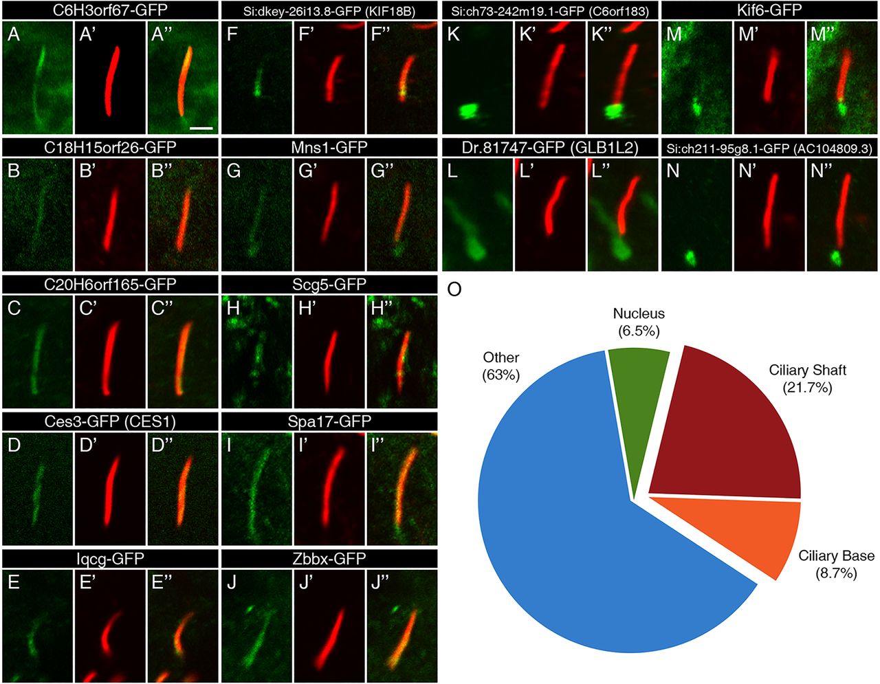
Fig. 2
Ciliary localization of FIG-encoded proteins in the zebrafish embryo. (A-N) The localization of GFP-tagged FIG-encoded proteins relative to motile cilia of the KV (green) in 13-14hpf embryos. The GFP signal was amplified with anti-GFP antibodies. (A2-N2) Ciliary axonemes were labeled with anti-acetylated tubulin antibodies (red). (A3-N3) Overlays of the two channels show the overlap between the gene products and the ciliary shafts. Scale bar: 2μm. (O) An overview of the localization of the FIG gene products shows that the majority does not localize to the cilium. These genes may encode cytoplasmic, membrane or nuclear regulators of cilia function.