
IMAGE

Fig. S9

ID
ZDB-IMAGE-141023-18
Genes
Publication
Alexander et al., 2014 - Wnt signaling interacts with bmp and edn1 to regulate dorsal-ventral patterning and growth of the craniofacial skeleton
All Figures
Figures for Alexander et al., 2014
Image
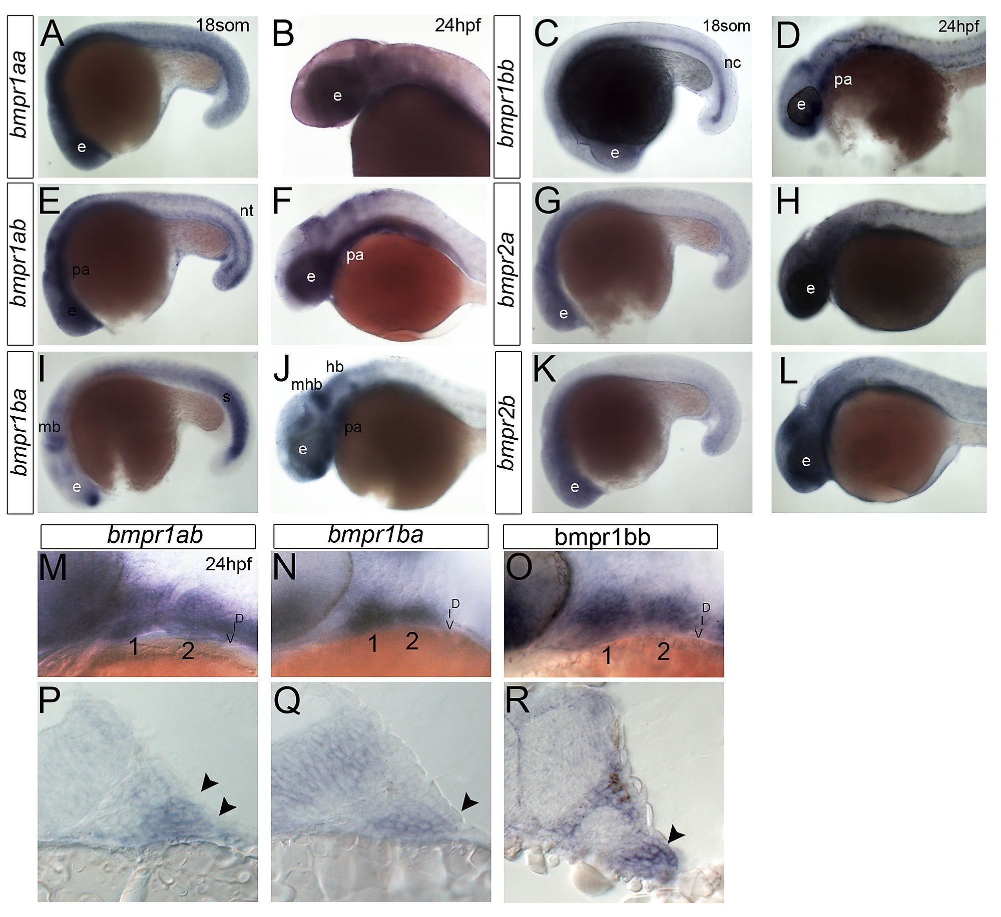
Figure Caption

Fig. S9

Bmp receptor expression during pharyngeal arch development. (A–O) Whole mount ISH for bmpr1aa (A, B), bmpr1bb (C, D, O), bmpr1ab (E, F, M), bmpr2a (G, H), bmpr1ba (I, J, N), and bmpr2b (K, L), lateral views, anterior to the left. (P–R) Transverse sections through 24 hpf embryos showing expression of bmpr1ab (P), bmpr1ba (Q), and bmpr1bb (R) in neural crest cells of the pharyngeal arches. Arrowheads indicate ventral restriction of bmpr1ab (P) and bmpr1bb (Q) expression compared with bmpr1ab (R). Abbreviations: e, eye; hb, hindbrain; mhb, mid-hindbrain boundary; nc, notocord; nt, neural tube; pa, pharyngeal arches; s, somites.

Figure Data
Acknowledgments
This image is the copyrighted work of the attributed author or publisher, and ZFIN has permission only to display this image to its users. Additional permissions should be obtained from the applicable author or publisher of the image. Full text @ PLoS Genet.