
Fig. 2
- ID
- ZDB-IMAGE-141016-22
- Publication
- Zdebik et al., 2013 - Epilepsy in kcnj10 Morphant Zebrafish Assessed with a Novel Method for Long-Term EEG Recordings
- All Figures
- Figures for Zdebik et al., 2013
|
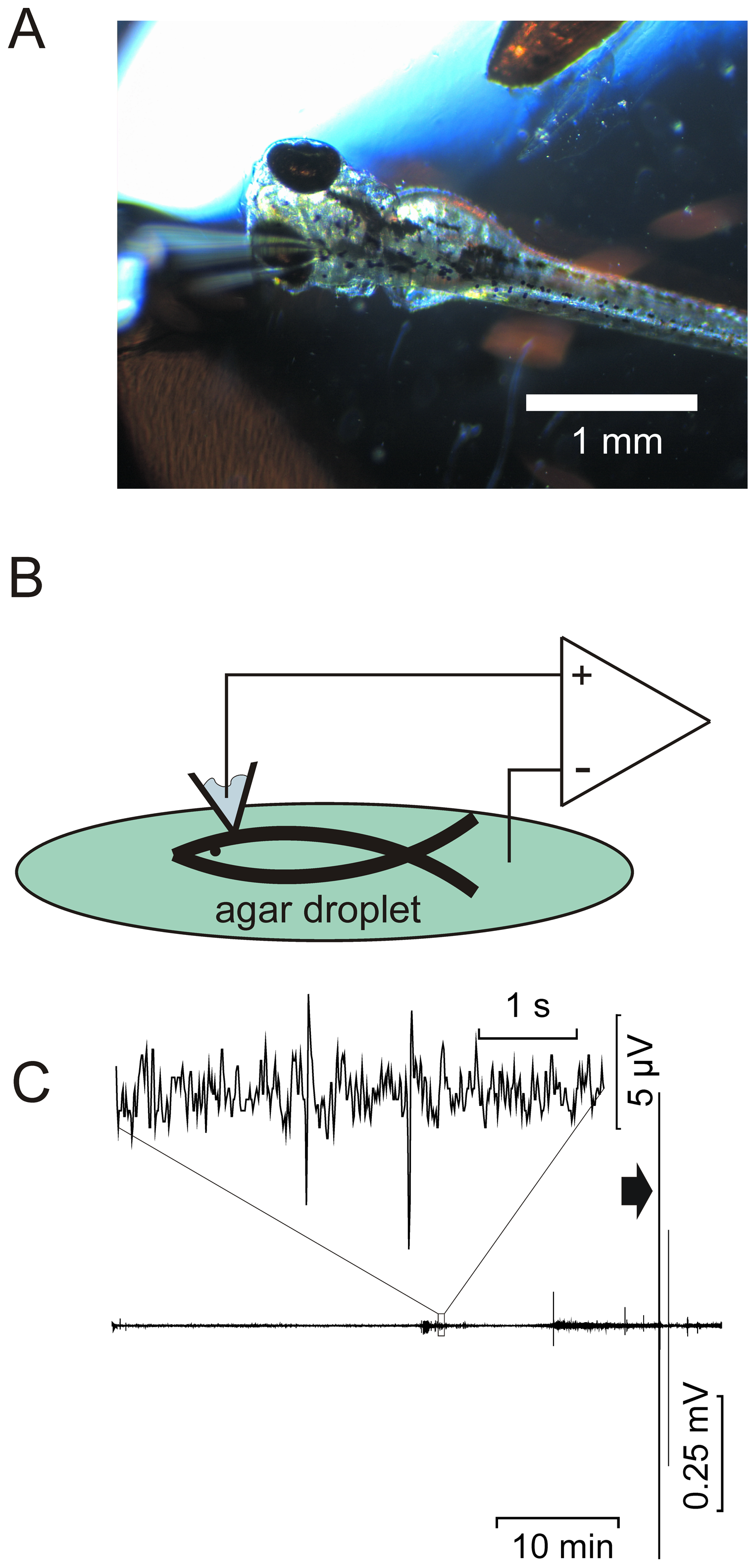
Fig. 2
Non-invasive recordings using a patch pipette on the surface of the optic tectum.
(A) Photo of the non-invasive EEG ZF recording set-up. To the left is a surface recording pipette, filled with 1 M NaCl. In the upper right is the reference electrode. Both are connected to the amplifier, as shown in the schematic in (B). The mounted fish is positioned on a microscope with which ZF viability can be monitored continuously. (C), long-term EEG recording of a kcnj10a morphant fish initially paralyzed in 20 mM D-tubocurarine. Fish were generally viable for over an hour, but paralysis appeared to wear off after 50 to 60 min. Movement artifacts ensued, associated with electrical activity and visible twitching (arrowhead). A 5 s period indicated by a box in C is represented above the trace in higher resolution.