Fig. 1
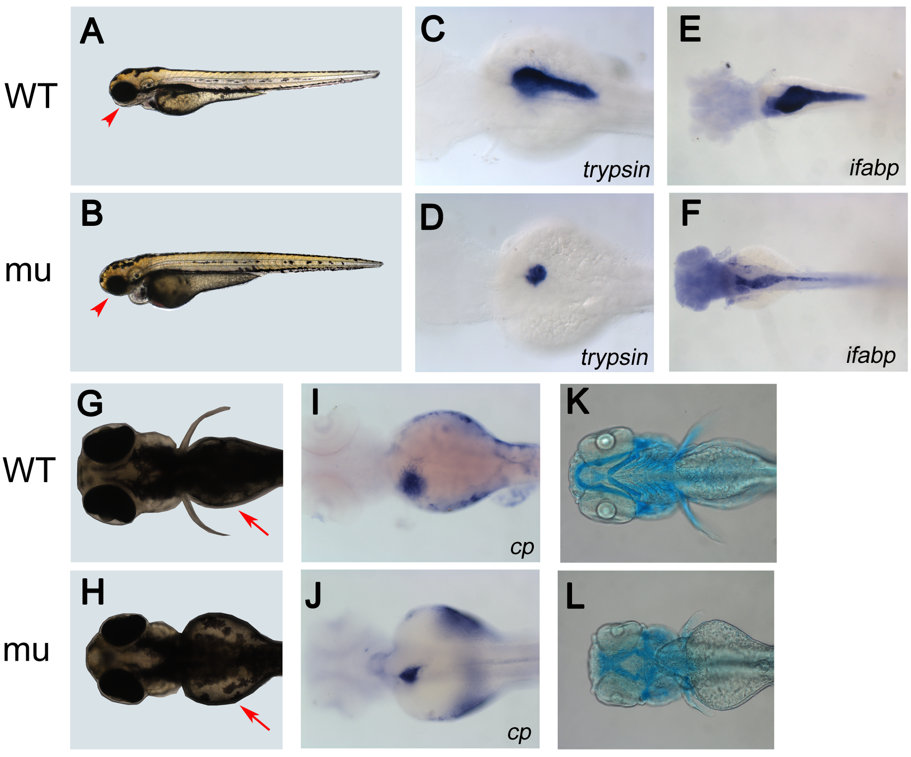
dg5 mutant has an endodermal and craniofacial defect.
(A, B) Lateral and (G, H) dorsal views of live Wild Type (WT) sibling and dg5 larvae at 3 dpf. Smaller head and eyes can be seen (arrowhead in A, B). Impaired yolk absorption is apparent in dg5 mutant at 3 dpf (arrows in G, H). (C–F, I–L) All dorsal views, anterior to the left. At 3 dpf, the expression of trypsin (try) is markedly reduced in dg5 mutant (D) as compared to WT group (C). The intestine marker fatty acid binding protein 2 (ifabp) expression reveals that the intestine is thinner in dg5 larvae (E) as compared to WT group (F) at 3.5 dpf. The expression of ceruloplasmin (cp) shows that the liver is smaller in dg5 mutant (H) as compared to WT group (I) at 3 dpf. As compared to control group (K), craniofacial development is abnormal in dg5 larvae (L) by alcian blue staining at 4 dpf.

