
Fig. 4
- ID
- ZDB-IMAGE-140916-1
- Genes
- Publication
- Venkatesh et al., 2014 - Elephant shark genome provides unique insights into gnathostome evolution
- All Figures
- Figures for Venkatesh et al., 2014
|
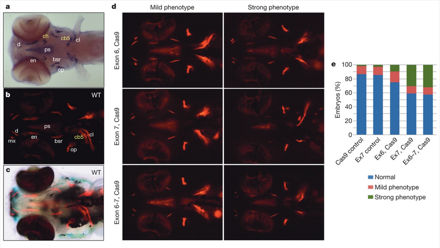
Fig. 4
a, spp1 is specifically expressed in cells surrounding the bone matrix. Ventral view of a 5-dpf embryo hybridized with a spp1-specific RNA probe. Yellow labels, endochondral bones (cb5, ceratobranchial5; ch, ceratohyal); white labels, dermal bones (bsr, branchiostegal ray; cl, cleithrum; d, dentary; en, entopterygoid; op, operculum; ps, parasphenoid). b, Ventral view of a 5-dpf wild-type (WT) embryo stained with alizarin red to reveal sites of bone deposition (red fluorescence). mx, maxilla. c, Bright-field image merged with b to visualize anatomical structures and locations of bone deposition simultaneously. d, Ventral views of 5-dpf embryos injected with Cas9 mRNA together with single guide RNA (sgRNA) targeting spp1 exon6, exon7 or both (alizarin red staining). The embryos were scored as normal (resembling wild type), mild or strong bone phenotypes, with the latter showing the greatest reduction in bone formation. The variations in the extent of bone reduction are presumably due to somatic chimaerism with regard to spp1 disruption. e, Proportions of mild and strong bone phenotypes induced by disruption of spp1 by sgRNA/Cas9. Targeting of exon7 (n = 206 embryos) or both exons6 and 7 (n = 143) resulted in significantly higher proportions of strong bone phenotype (P<0.01, Fisher’s exact test) compared with control injections of Cas9 mRNA (n = 190) and exon7 sgRNA (n = 143) (Ex6, Cas9: n = 72).