
Image
|
Figure Caption
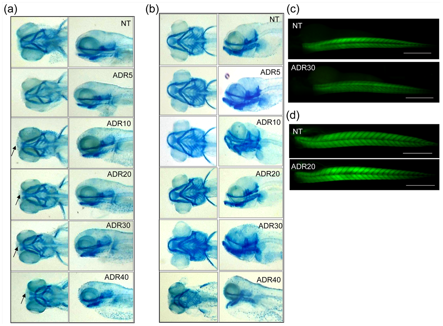
Fig. 9
Cartilage development.
At 72(a), the craniofacial cartilage, detected by alcian blue staining, shows a delay of development (arrows) in fish exposed to 10–40 μM adriamycin. (b) At 120 hpf, larvae exposed to adriamycin up to 30 μM are indistinguishable from the control in terms of craniofacial morphology. Instead, exposure to adriamycin 40 μM causes persistent alteration of the cartilage structure. FITC-phalloidin staining shows normal muscle morphology of 72 hpf (c) and 120 hpf (d) larvae exposed to 20 μM adriamycin.
Figure Data
Acknowledgments
This image is the copyrighted work of the attributed author or publisher, and
ZFIN has permission only to display this image to its users.
Additional permissions should be obtained from the applicable author or publisher of the image.
Full text @ PLoS One