
IMAGE
Fig. 2
- ID
- ZDB-IMAGE-140821-4
- Genes
- Publication
- Loh et al., 2014 - Zebrafish yap1 plays a role in differentiation of hair cells in posterior lateral line
- All Figures
- Figures for Loh et al., 2014
Image
|
Figure Caption
Fig. 2
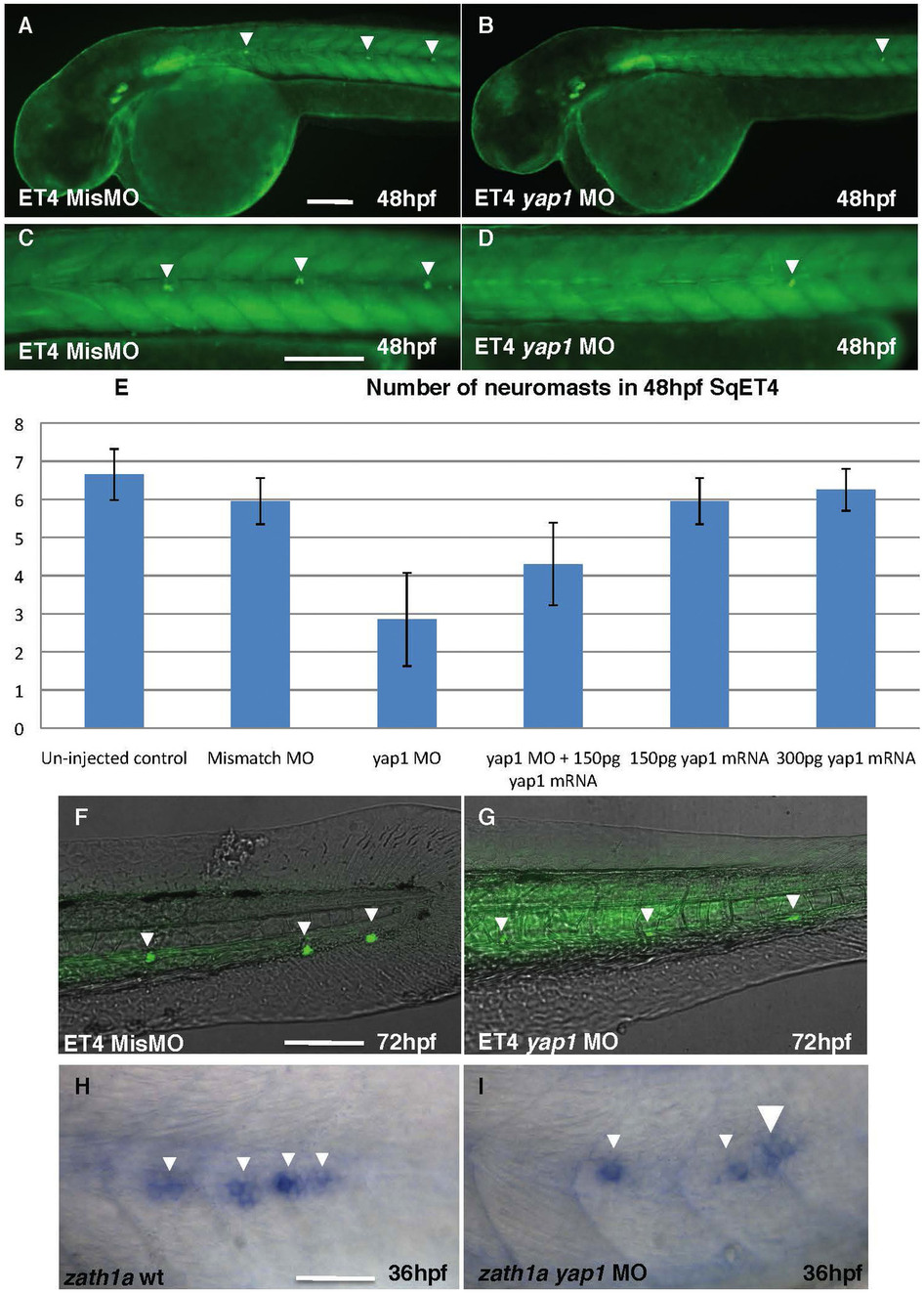
(A), (C), (F) – controls; (B), (D), (G) – Yap1 morphants. All embryos are shown in lateral view. The table illustrates the rescue effect of yap1 mRNA coinjection. The original data are provided as a supplement. (A), (B) – head and anterior trunk, (C), (D) – intermediate trunk, (F), (G) – tail. Notice a reduction in a number of neuromast cells and increase of background due to longer exposure in (G) comparing to (F). (H), (I) – zath1a expression in the lateral line primordium of controls (H) and morphants (I). Scale bar = 40 μm, (H)&(J) = 20 μm.
Figure Data
Acknowledgments
This image is the copyrighted work of the attributed author or publisher, and
ZFIN has permission only to display this image to its users.
Additional permissions should be obtained from the applicable author or publisher of the image.
Full text @ Sci. Rep.