
|
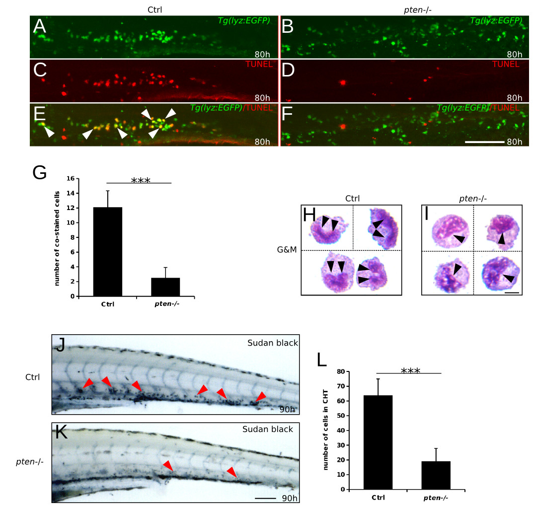
Fig. 2
Reduced apoptosis and block in myeloid cell maturation underlie pten deficiency-induced expansive myelopoiesis. (A-G) Double staining of lyz-driven EGFP protein and TUNEL assay in the CHT of control and pten-/- embryos at 80 hpf. Although relative more EGFP-positive myeloid cells were observed in the pten-/- embryos (A and B, all green cells were manually counted as positive), few cells were simultaneously apoptotic, as indicated by merging with TUNEL staining (E and F, white arrowheads). (H and I) Wright-Giemsa staining and morphological characterization of FACS-purified EGFP-positive cells from 90 hpf pten-/-lyz:EGFP and control embryos. The black arrowheads indicate the nuclear shape, which was typically kidney-like and condensed in control embryos (H) but remained in a loose state in the pten-/- embryos (I). (J-L) Sudan Black staining of neutrophils at 90 hpf. The red arrowheads indicate neutrophils in the CHT. The data shown in (G) and (L) are the means ± SEM of at least 15 and 30 embryos; ***p < 0.001 versus the control. Scale bar: h and i, 5 um; others, 100 μm.