
Fig. 3
- ID
- ZDB-IMAGE-140812-19
- Antibodies
- Publication
- Wiweger et al., 2014 - Possible effects of EXT2 on mesenchymal differentiation--lessons from the zebrafish
- All Figures
- Figures for Wiweger et al., 2014
|
Fig. 3
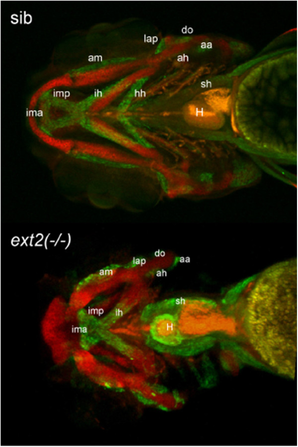
Homozygous ext2 mutant displays musco-skeletal phenotype. Whole mount immunolocalisation at 4dpf using MF-20 antibody for muscles (green) and collagen II for cartilage (red) shows thicker and shorter muscles fitting the malformed cartilaginous skeleton in the ext2-/- fish. Muscles: intermandibularis anterior (ima), intermandibularis posterior (imp), adductor mandibulae (am), interhyoideus (ih), hyohyoideus (hh), levator arcus palatine (lap), adductor hyoideus (ah), dilator operculi (do), sternohyoideus (sh) and adductor operculi (ao). Note missing hh muscle and, marked with a star, increased musculature around heart (H) in ext2-/-. Scale = 0.1 mm.