
Image
|
Figure Caption
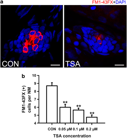
Fig. 2
Detection of neuromast hair cells with live staining (FM1-43FX) in 5dpf larvae. (a) Confocal images of neuromasts from a control and 0.1μM TSA-treated larva at 5dpf that have been labeled for FM1-43FX to detect functional HCs. Fewer FM1-43FX-positive cells developed in the TSA-treated larva compared with the untreated control. Nuclei are stained with DAPI and scale bars=10μm. (b) The average number of functional HCs per neuromast (NM) in 5dpf zebrafish larvae following TSA treatment. **P<0.001.
Acknowledgments
This image is the copyrighted work of the attributed author or publisher, and
ZFIN has permission only to display this image to its users.
Additional permissions should be obtained from the applicable author or publisher of the image.
Full text @ Exp. Mol. Med.