
|
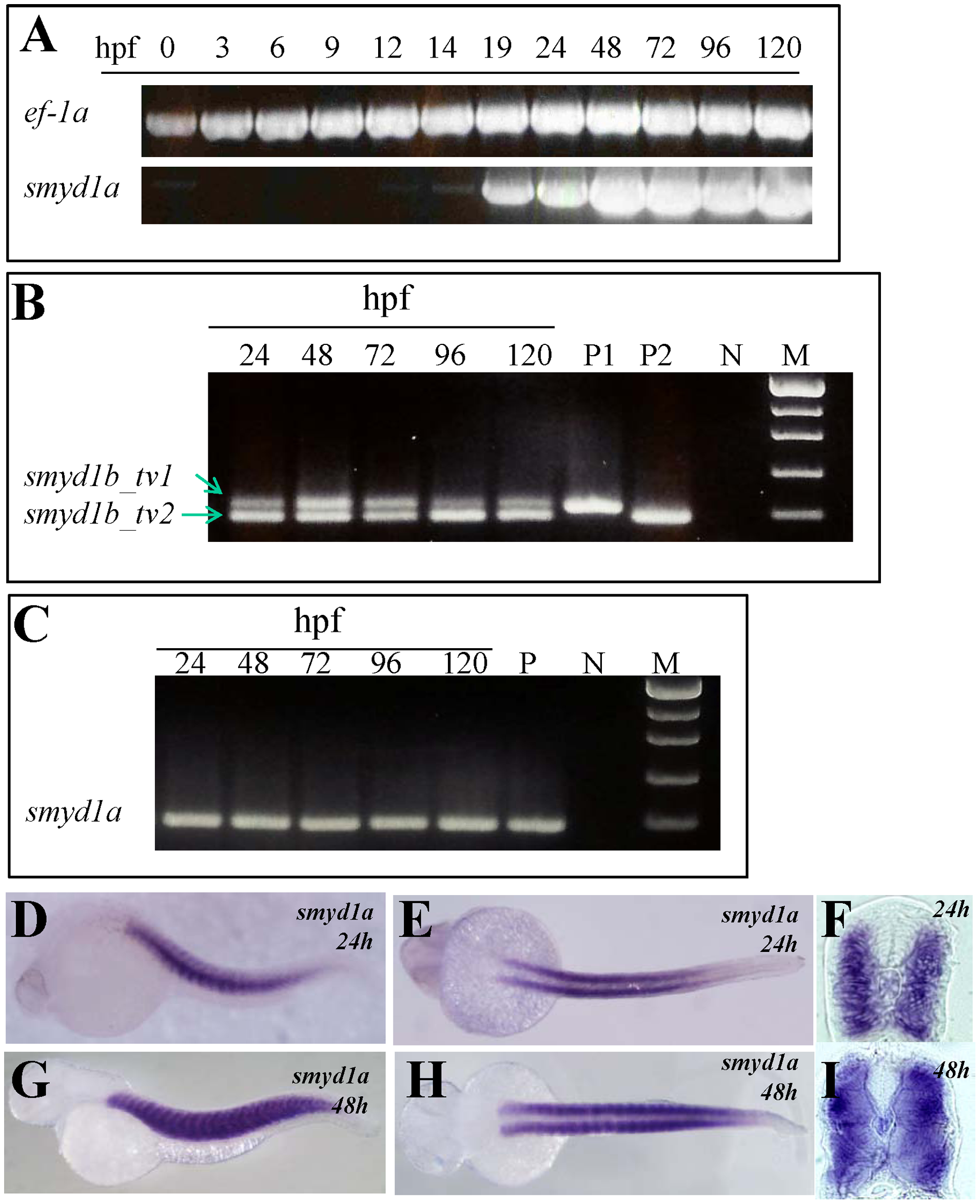
Fig. 1
The temporal and spatial pattern of smyd1a expression in zebrafish embryos.
A. RT-PCR results show temporal expression of smyd1a in zebrafish embryos from fertilization to day 5. Elongation factor 1-alpha (ef-1α) was used as control. B. RT-PCR analysis shows the alternative splicing of smyd1b exon 5 generating two isoforms of smyd1b, smyd1b_tv1 and smyd1b_tv2. C. RT-PCR analysis shows the lack of alternative splicing of exon 5 in smyd1a in zebrafish embryos. D–I. Whole mount in situ hybridization shows the spatial pattern of smyd1a mRNA expression using a dig-labeled antisense probe. smyd1a expression was detected in skeletal muscles of zebrafish embryos at 24 (D, E, F) and 48 (G, H. I) hpf. D, G represent the side view; E, H repre4sent the dorsal view; F, I represent the cross sections.