
Fig. 3
- ID
- ZDB-IMAGE-140804-14
- Genes
- Antibodies
- Publication
- Elks et al., 2014 - Mycobacteria Counteract a TLR-Mediated Nitrosative Defense Mechanism in a Zebrafish Infection Model
- All Figures
- Figures for Elks et al., 2014
|
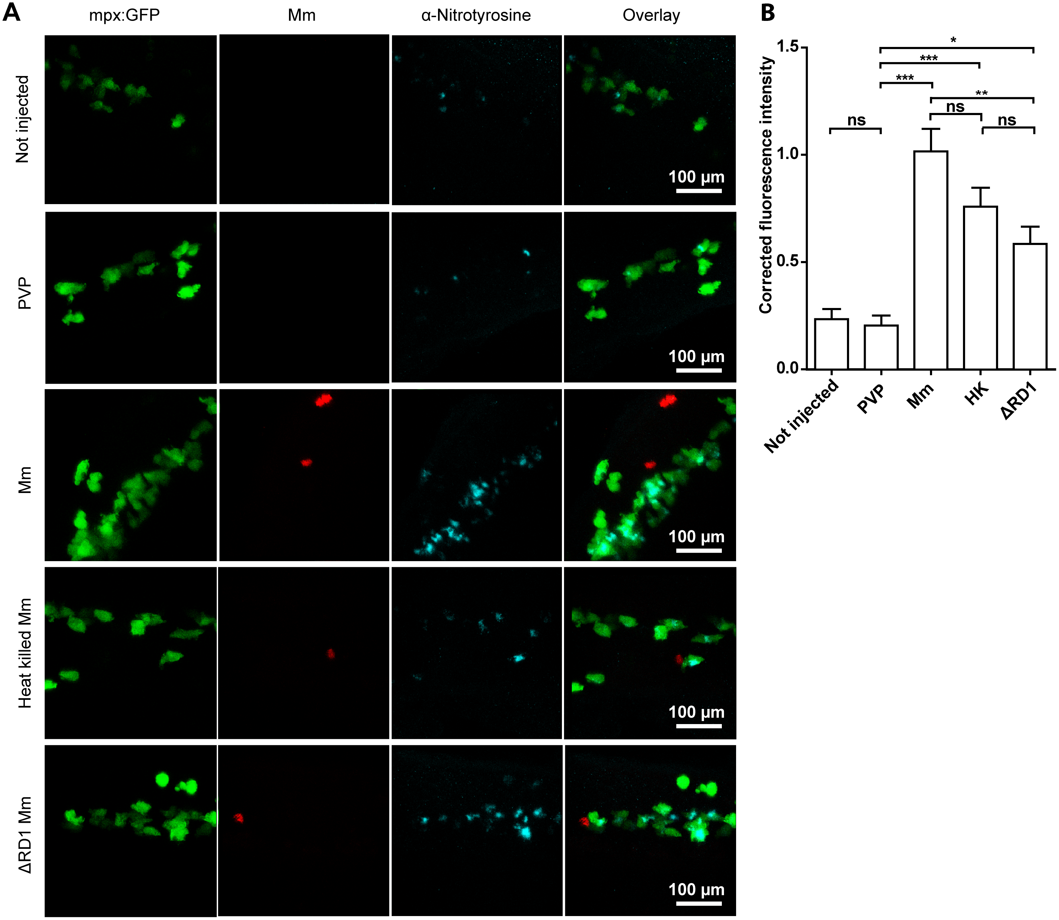
Fig. 3
Injection with live, heat-killed or ΔRD1 Mm increased tyrosine nitration levels.
(A) Example fluorescence confocal z-stacks of the caudal vein region of embryos stained with anti-nitrotyrosine antibody, imaged 1 day after injection with live, heat killed or ΔRD1 Mm in comparison with uninjected embryos or embryos injected with 2% PVP carrier solution. (B) Corrected fluorescence intensity levels of anti-nitrotyrosine antibody confocal z-stacks of equal size 1 day after injection with live, heat killed or ΔRD1 Mm. Data shown are mean ± SEM, n = 58-99 cells from 15 embryos combined from 3 independent experiments.