
|
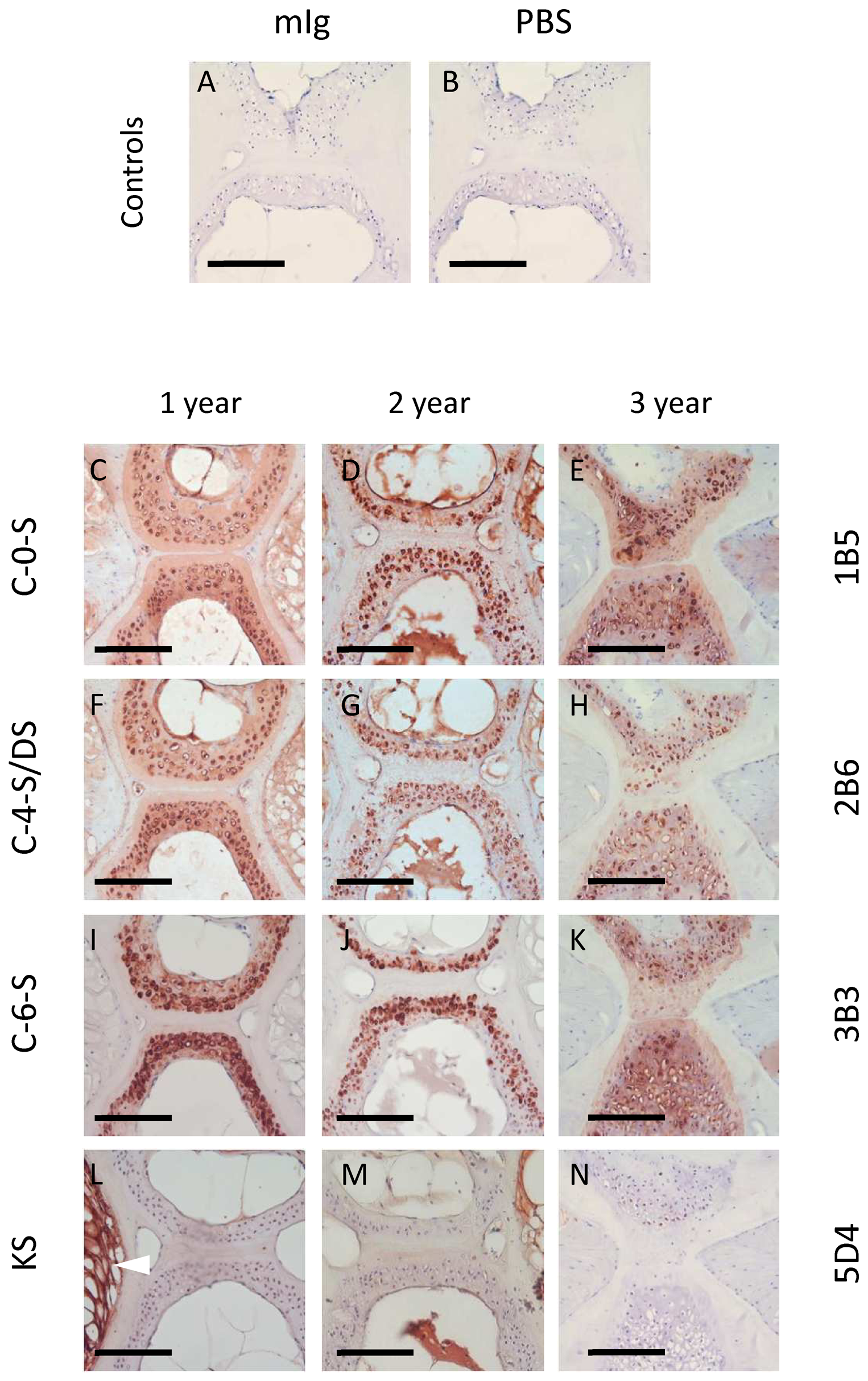
Fig. 4
The vertebral cartilage of aging fish is rich in chondroitin, but not keratan, sulfate.
A-B. Immunohistochemical labelling controls showing no non-specific binding of primary (mIg, ‘naive’ mouse immunoglobulin) or secondary antibody (PBS, phosphate buffered saline). C-N. Immunohistochemical labelling patterns of chondroitin/dermatan (C-0-S, C-4-S/DS, C-6-S) and keratan sulfate (KS) at 1, 2 and 3 years (left, middle and right panels, respectively). Note prominent pericellular labelling of CS/DS epitopes, particularly in 2 and 3 year samples. Unlike CS, KS occurs only within the notochordal tissue of the intervertebral disc (bottom left; arrowhead) and appears to diminish during aging. Scalebar represents 100 microns.