
Fig. 7
- ID
- ZDB-IMAGE-140723-59
- Genes
- Antibodies
- Publication
- Rydeen et al., 2014 - Cyp26 enzymes are required to balance the cardiac and vascular lineages within the anterior lateral plate mesoderm
- All Figures
- Figures for Rydeen et al., 2014
|
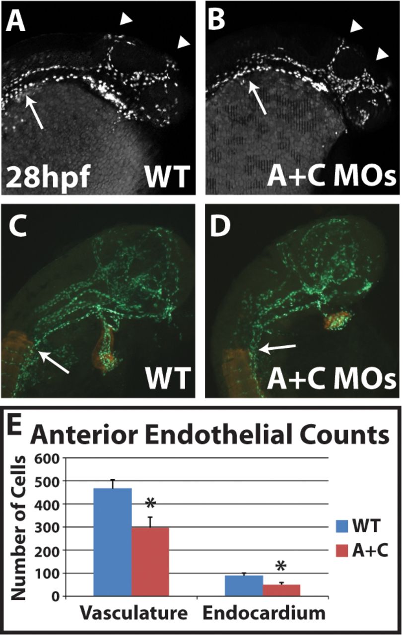
Fig. 7
Cyp26-deficient embryos have decreased anterior endothelial cell number. (A,B) Optical sections of uninjected sibling and Cyp26-deficient Tg(kdrl:nEGFP) embryos show that Cyp26-deficient embryos have mispatterned cranial vasculature. (C,D) Representative immunostained Tg(kdrl:nEGFP) embryos for counting the cranial endothelial and endocardial cells (green). Somites and myocardial cells are indicated in red. (E) Endothelial and endocardial cell counts at 28hpf. WT, n=10; Cyp26-deficient, n=11. All images are lateral views with anterior towards the right. Arrowheads in A and B indicate primordial midbrain channel and the anterior and middle cerebral veins. Arrows in A-D indicate the somite boundary. Significant differences compared with controls are indicated (*P<0.05). Error bars indicate s.d.