
|
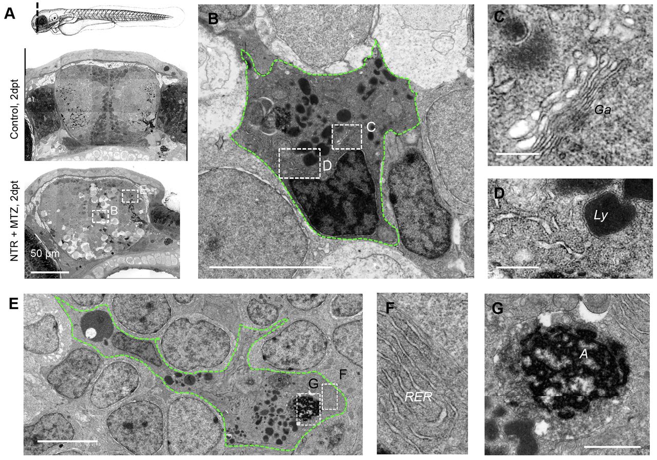
Fig. 4
Phagocytes with ultrastructural microglial features appear following neuronal cell death. (A) Nanotomy of brains of 7 dpf control and NTR animals treated with MTZ 2 days post treatment, showing features specific to the NTR degenerative brain, including phagocytic leukocytes, dark cells undergoing cell death and spongy appearance of neural tissue in comparison with control. (B–D) High magnification view of phagocytic cell in A (lower panel) showing typical amoeboid microglial features including prominent Golgi apparatus (Ga; C), inclusions including lysosomal vacuoles (Ly; D) and distinctive long stretches of endoplasmic reticulum (D). (E–G) High magnification view of phagocytic cell in A showing typical amoeboid microglial cell features, including rough endoplasmic reticulum (RER; F) and engulfed cell corpse (‘A’; G). Scale bars: 50 μm (A), 200 nm (C,D), 5 μm (B,E), 1 μm (G).