
IMAGE
Fig. 8
- ID
- ZDB-IMAGE-140707-11
- Publication
- Dash et al., 2014 - sept7b is essential for pronephric function and development of left-right asymmetry in zebrafish embryogenesis
- All Figures
- Figures for Dash et al., 2014
Image
|
Figure Caption
Fig. 8
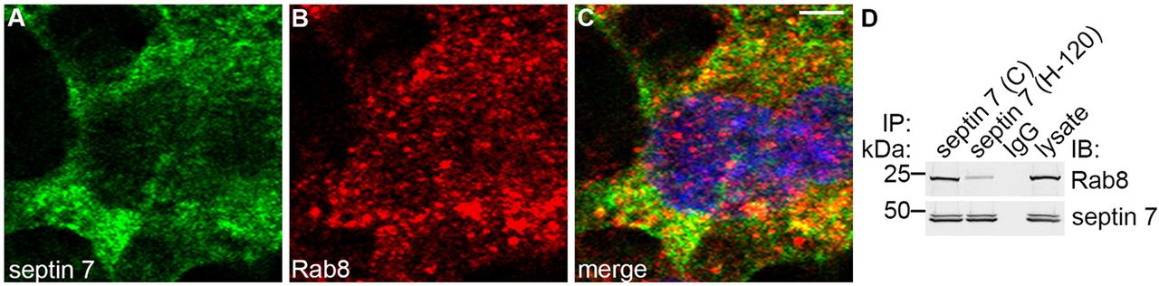
Septin 7 forms a complex with Rab8. (A–C) Immunofluorescence staining of mIMCD3 cells shows partial colocalization of septin 7 (A) and Rab8 (B) in the perinuclear region, as shown by the yellow coloring in the merged image (C). Scale bar: 10μm. (D) Two different septin 7 antibodies (C or H-120) co-immunoprecipitate Rab8 in mIMCD3 cell lysates, indicating that septin 7 and Rab8 physically interact. IP, immunoprecipitation; IB, immunoblotting.
Acknowledgments
This image is the copyrighted work of the attributed author or publisher, and
ZFIN has permission only to display this image to its users.
Additional permissions should be obtained from the applicable author or publisher of the image.
Full text @ J. Cell Sci.