
Fig. S4
- ID
- ZDB-IMAGE-140703-3
- Genes
- Publication
- Leitch et al., 2014 - Basal body proteins regulate Notch signaling via endosomal trafficking
- All Figures
- Figures for Leitch et al., 2014
|
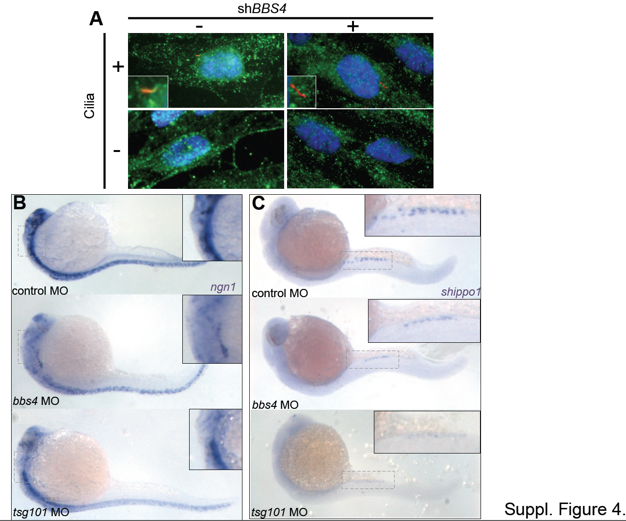
Fig. S4
Notch1 localization in ciliated and unciliated cells and suppression of Notch-inhibited cell type markers in bbs4 morphants. (A) hTERT-RPE1 cells doubled immunostained for detection of NOTCH1 (green) and ARL13B (red) and transfected with or without shBBS4. 40X magnification. Cilia in ciliated cells (+) magnified in inset boxes. Scale bars = 20 μm. (B) Expression of neurogenin1 in control MO-, bbs4 MO- and tsg101 MO-injected embryos at 24 hpf detected by whole mount in situ hybridization. (C) Expression of shippo1 in control MO-, bbs4 MO- and tsg101 MO-injected embryos at 24 hpf. Insets indicate magnification of dashed boxes.