
|
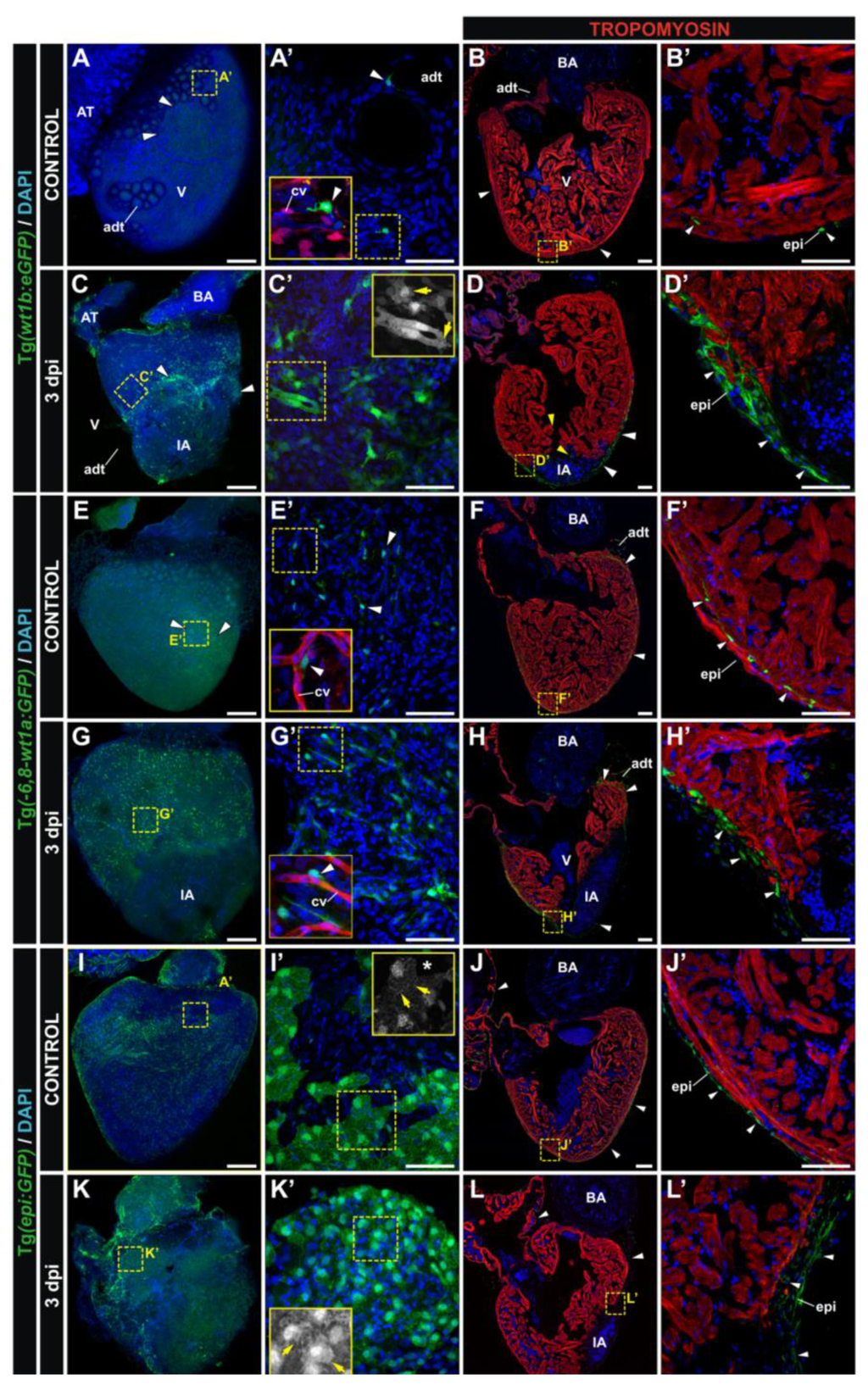
Fig. 4
Transgenic reporter lines that mark the adult epicardium during regeneration.Whole mount confocal 3D projections and immunofluorescence on sections of dissected control and cryoinjured hearts at 3 days post-injury (dpi). Hearts are from the Tg(wt1b:GFP) (A-D), Tg(-6.8kbwt1a:GFP) (E-H) and Tg(epi:GFP) (I-L) lines. Anterior is to the top; ventral to the right. Nuclei are counterstained with DAPI (blue). (A′-L′) Zoomed images of the boxed areas in A-L. Insets in (A′), (E′) and (G′) additionally show details of the coronary vasculature (red), reported by the Tg(fli1a:DsRedEx) transgene. Insets in (C′), (I′) and (K′) show the GFP channel from the boxed area. White arrowheads mark GFP-positive cells; yellow arrows mark morphological cell features. adt, adipose tissue; AT, atrium; BA, bulbus arteriosus; cv, coronary vessels; epi, epicardium; IA, injured area; V, ventricle. Bars: 200 µm (full views), 50 µm (magnifications).