
|
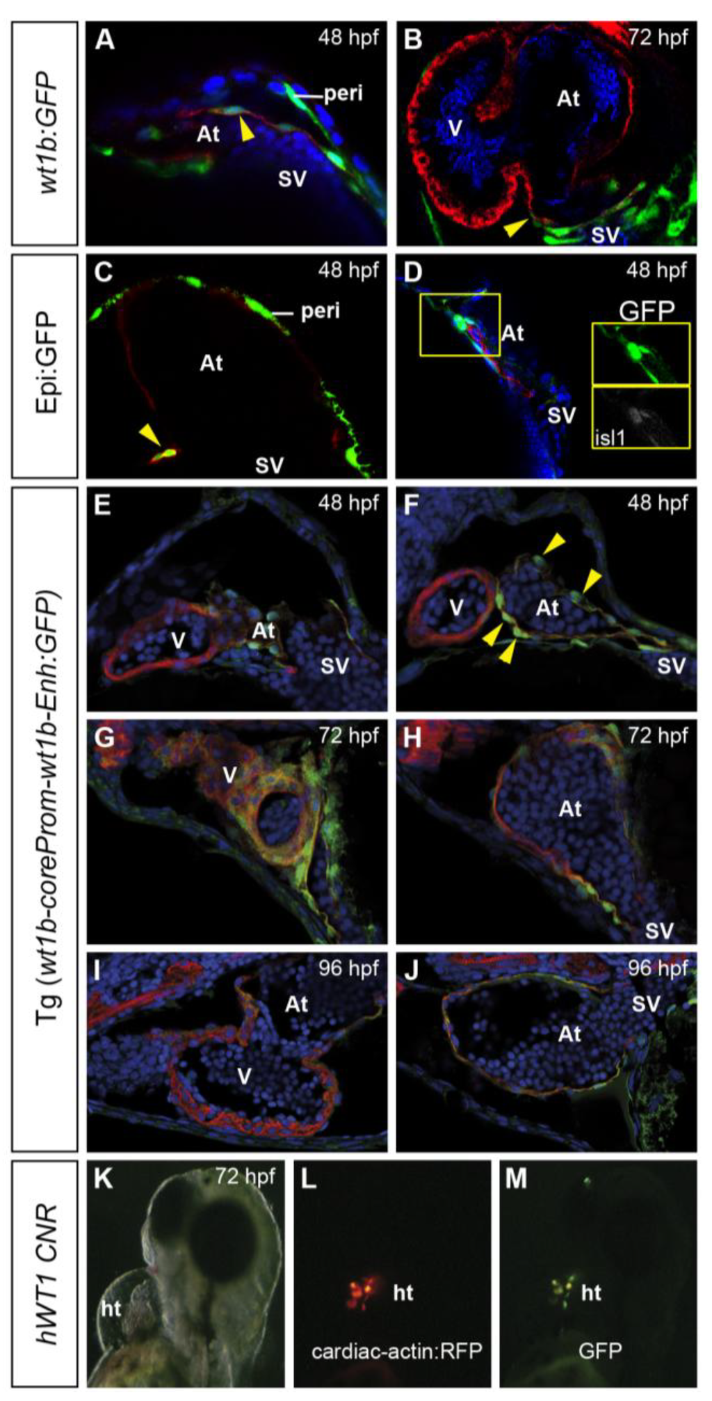
Fig. 3 Partial coexpression of GFP and myocardial marker genes in wt1 transgenic lines. Immunofluorescence on sections of different wt1 transgenic lines at stages are indicated in the panels, revealing GFP expression in green and myosin heavy chain (MHC) expression in red, except in panel A, in which red reveals myh6 expression. Cell nuclei are counterstained in blue. Arrowheads mark cells coexpressing GFP and a cardiomyocyte marker. (A and B) wt1b:GFP at 48 and 72 hpf, showing GFP-positive myocardial cells close to the venous pole. (C) Coexpression of Epi:GFP with myh6 in atrial cardiomyocytes close to the venous pole. (D) Coexpression of Epi:GFP with islet 1 (isl1) at the venous pole of the heart. (E–J) Tg(wt1b-coreProm-wt1b-Enh:GFP) embryos expressing GFP in the atrial myocardium. (K–M) Brightfield and fluorescence views of a transient transgenic larva expressing GFP under the control of a conserved human WT1 noncoding genomic region (CNR) and expressing red fluorescent protein (RFP) under the control of a myocardial promoter. Note the cells coexpressing GFP and RFP. At, atrium; ht, heart tube; hpf, hours postfertilization; peri, pericardial mesothelium; SV, sinus venosus; V, ventricle.