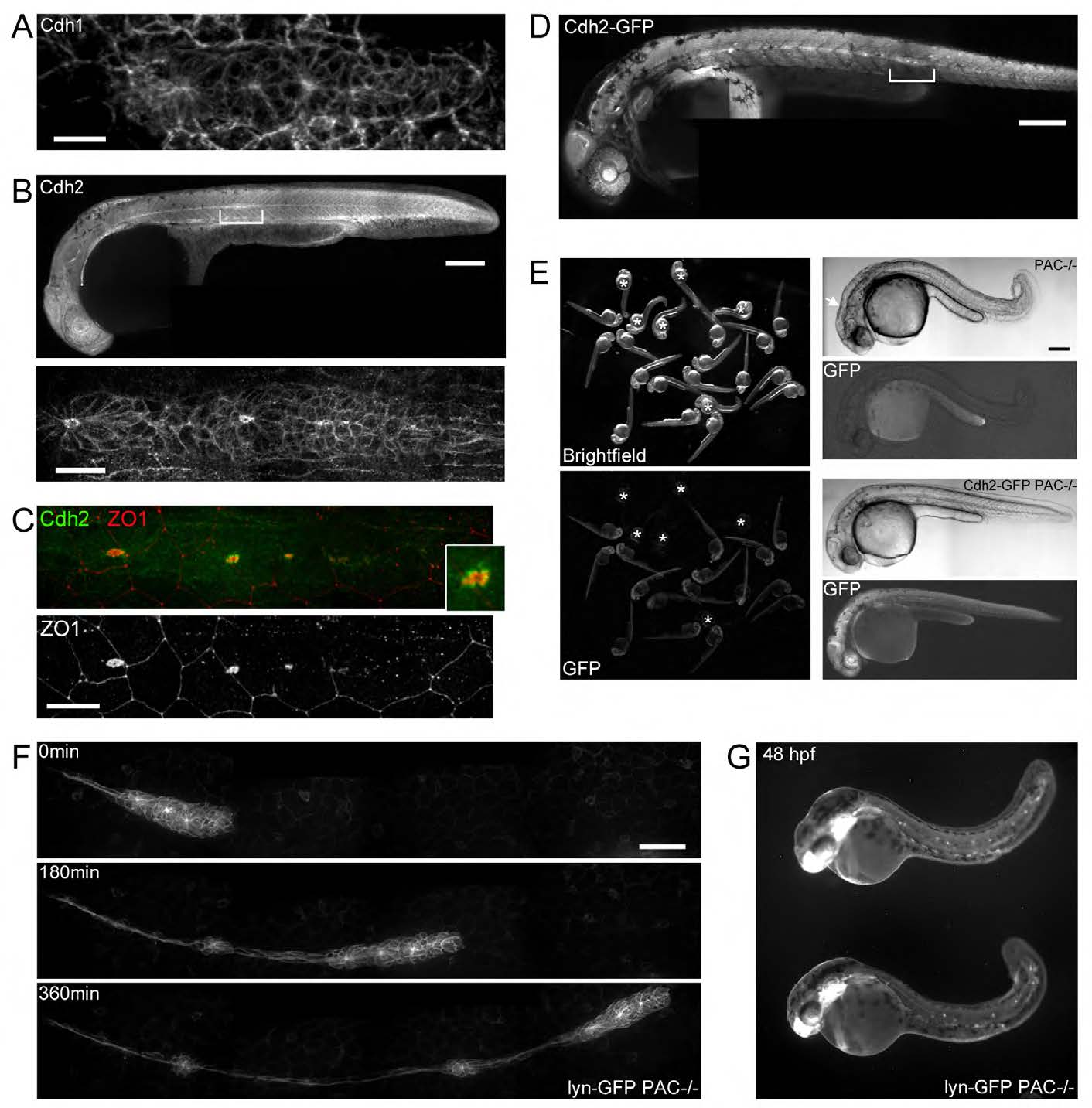
Fig. S2 Generation of live adherens junction reporter lines (A) Immunohistochemistry of Cdh1 in the pLLP. (B) Immunohistochemistry of Cdh2 in a 30hpf embryo and in the pLLP (lower panel). (C) Double antibody staining for Cdh2 and ZO1 in the pLLP. The twice-enlarged view of a rosette highlights the broader localisation of Cdh2 around ZO1. (D) Whole embryo overview of the cadherin2:Cadherin2-GFP BAC that recapitulates endogenous Cdh2 expression. Brackets highlight the position of the pLLP. (E) Rescue of the parachute mutant (cdh2tm101b-/-, PAC-/-) by cadherin2:Cadherin2-GFP transgenics. Left panels show a clutch of embryos from an incross of cadherin2:Cadherin2-GFP;cdh2tm101b-/-. Non-transgenic embryos (*) show characteristic mutant phenotype illustrated in the top right panels (curled tail and brain defects, white arrow) whereas transgenic siblings develop normally (bottom right) and survive. Scale bars embryo=200µm, pLLP=20µm. (F) Images from a time-lapse movie of a cldnb:lyn-GFP;cdh2tm101b-/- showing the migration of the pLLP at 30hpf and the formation and deposition of neuromasts in the absence of Cdh2. Scale bar=50µm.(G) 48hpf cldnb:lyn-GFP;cdh2tm101b-/- embryos depicting the posterior lateral line neuromast pattern after the pLLP has reached the tail.
Image
Figure Caption
Acknowledgments
This image is the copyrighted work of the attributed author or publisher, and
ZFIN has permission only to display this image to its users.
Additional permissions should be obtained from the applicable author or publisher of the image.
Full text @ Development

