
Fig. S1
- ID
- ZDB-IMAGE-140416-18
- Publication
- Elks et al., 2013 - Hypoxia inducible factor signaling modulates susceptibility to mycobacterial infection via a nitric oxide dependent mechanism
- All Figures
- Figures for Elks et al., 2013
|
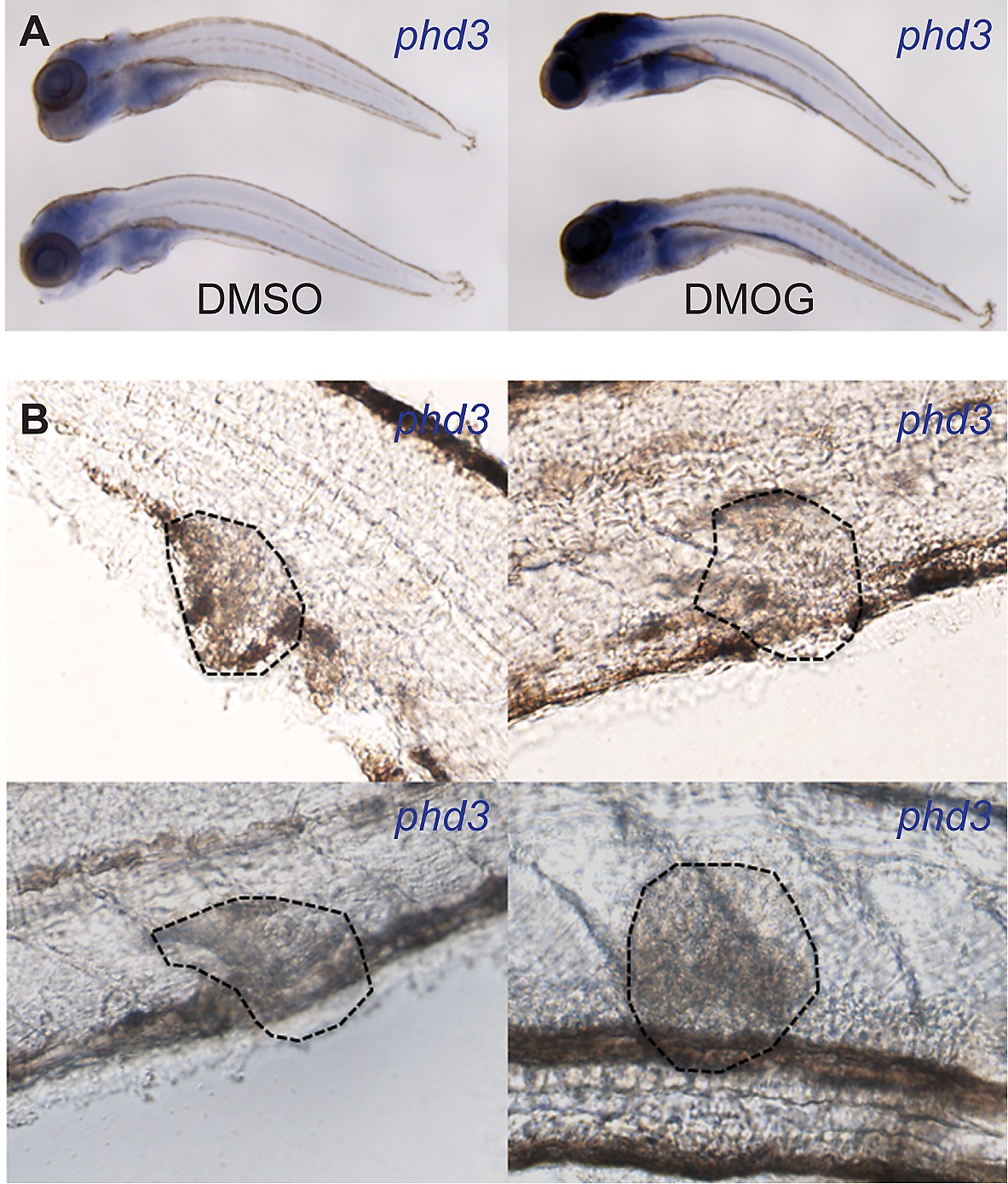
Fig. S1 Later stage larval infection showed no detectable levels of Hif-1α signaling. (A) In situ hybridization using a phd-3 antisense probe indicated no detectable expression in granulomas in 6 dpi zebrafish larvae. Lower panels show larvae treated with DMOG that have upregulated expression of phd3 indicating that the in situ detection of phd3 is functional and dependent on activated Hif-α signaling. (B) Micrographs of 4 individual granulomas (encircled with dotted lines) of 6 dpi larvae taken with DIC light microscopy. Hif-α signaling is labeled in the larvae by phd3 in situ hybridization, however, no levels of expression were detectable in granulomas.